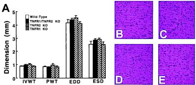
Figure 1.
Myocardial structure and histology. (A) Values (mm) for LV interventricular wall thickness (IVWT), posterior wall thickness (PWT), end-diastolic dimension (EDD), and end-systolic dimension (ESD) in the wild-type, TNFR1-deficient [TNFR1 knockout (KO)], TNFR2-deficient (TNFR2 KO), and TNFR1/TNFR2-deficient (TFR1/TNFR2 KO) mice, as measured by M-mode echocardiography. (B–E) Representative hematoxylin and eosin-stained myocardial sections of wild-type mice (B), TNFR1-deficient mice (C), TNFR2-deficient mice (D), and TNFR1/TNFR2-deficient mice (E) photographed at ×400.

