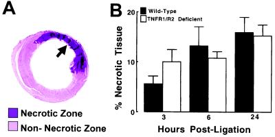
Figure 5.
Percentage of necrotic myocardium in wild-type and TNFR1/TNFR2-deficient mice. The area of necrosis within the area at risk was determined by planimetry of the myocardial sections that were labeled purple by the anti-myosin Ab, as shown by the arrow depicted in A. (B) Results of group data in wild-type and TNFR1/TNFR2-deficient mice. The extent of necrosis is expressed as the percentage of necrotic tissue in the LV myocardium (see Methods).

