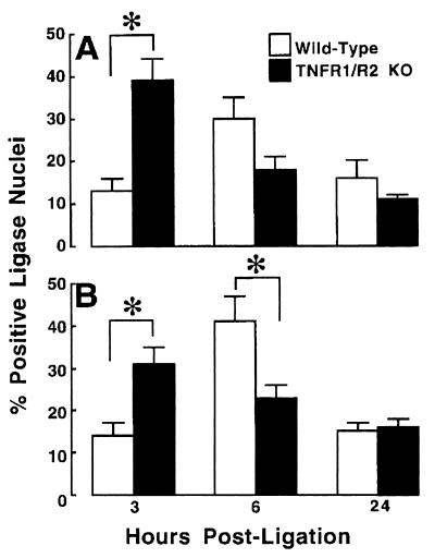
Figure 6.
Frequency of apoptosis in wild-type TNFR mice and TNFR1/TNFR2-deficient mice. The frequency of apoptosis was characterized with the necrotic zone (A) and the nonnecrotic zone (B) within the area at risk. The frequency of cardiac myocyte apoptosis was expressed as a total of the number of cardiac myocytes per 10,000 μm2 of myocardium. Apoptosis was determined by using a ligase-based method (see Methods). Animals were killed at 3, 6, and 24 h after acute ligation of the LAD. ∗ = P < 0.05; KO, knockout.

