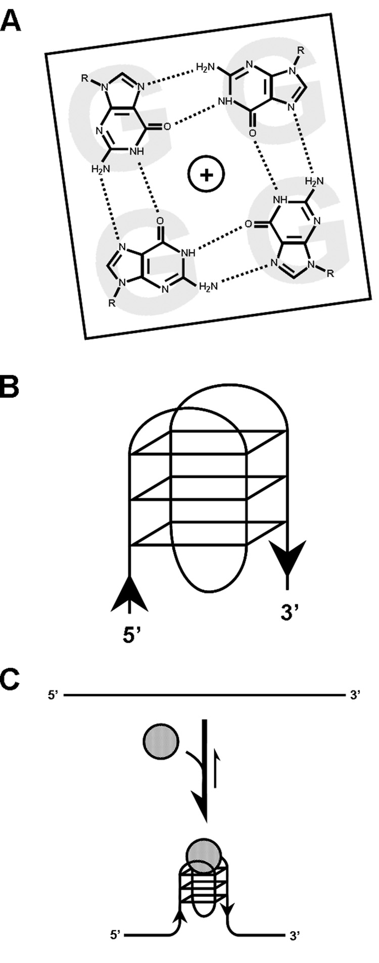
Figure 1.

G-quadruplex structure and ligand binding. (A) Structure of a G-quartet, with hydrogen bonds between guanines indicated by dotted lines. (B) Schematic of one type of unimolecular G-quadruplex that is composed of three planar G-quartet structures, as shown. The orientation (5’->3’) of the phosphodiester backbone is indicated by black arrows. Many other types of G-quadruplex folds are possible. (C) Binding of certain G-quadruplex interacting factors (grey circles), be they proteins or small molecular weight ligands, can promote and/or stabilize the formation of G-quadruplexes from nucleic acid strands with quadruplex-forming potential (QFP). Further, they might inhibit access of other factors to G-quadruplexes.
