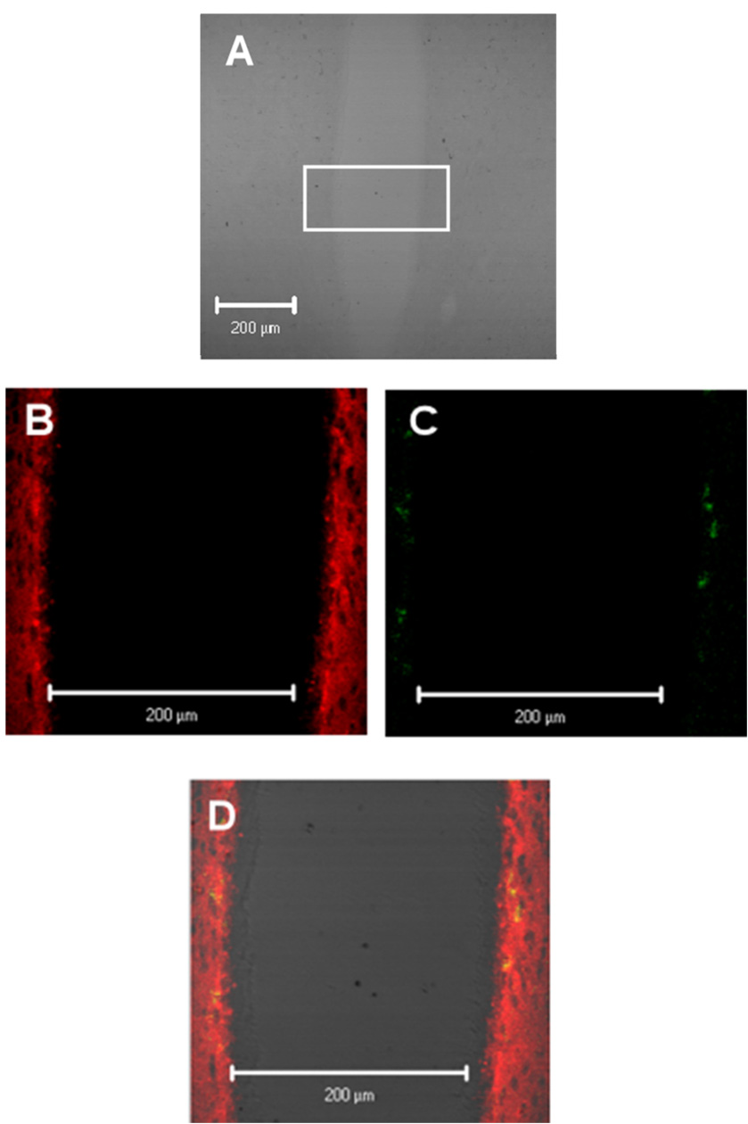
Figure 3.
IL-6 and IL-6R co-localization patterns in the 3rd ventricle region. Panel A is a 20x zoom of image 2B. Localization of IL-6 (red) and IL-6 receptor (IL-6R; green) are shown in Figure 3B and Figures 3B and 3C respectively. Overlay of figure 3B and figures 3C and 3C are shown in figure 3D with yellow representing co-localization of IL-6 and the IL-6R.

