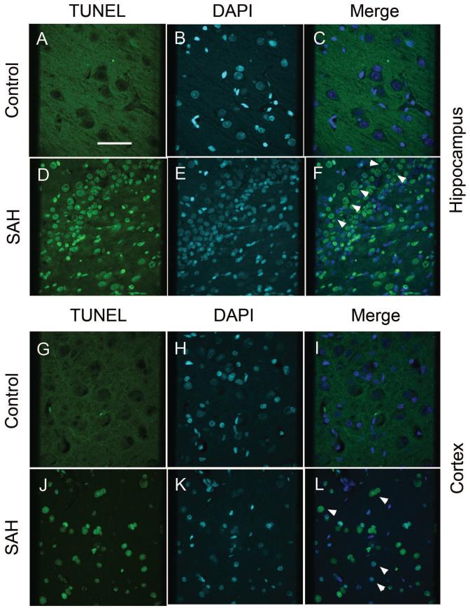
Figure 2. TUNEL staining in hippocampus and cortex of control and SAH dogs.
Images of TUNEL-positive (green) cells from control (A, G) and dogs with subarachnoid hemorrhage (SAH, D, J). Sections are from hippocampus (A-F) and cortex (G-L). There were no TUNEL-positive cells in control sections (A, G). However, in animals with SAH, cells stained positively for TUNEL (green, D, J) and co-localized with DAPI nuclear staining (white arrow heads, F, L). Scale bar = 150 μm.

