
Figure 1. Groove markers in wild-type embryos.
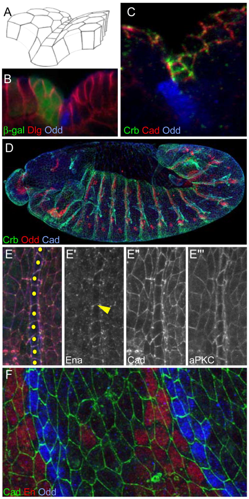
The figures are projections of confocal image stacks. Unless otherwise indicated, anterior is left and dorsal is up. Embryos are ~500μm long and cell diameters after stage 11 are ~4 μm.
(A) Schematic representation of the groove. Groove cells have rectangular junctions and are located at the bottom of the groove.
(B) Cross section of a stage 14 embryo expressing β-gal (green) in the en domain. Odd (blue) marks groove cells and Dlg (red) reveals cell shapes.
(C) Projection oriented as in (A) to display Crb accumulation (green) at the subapical domain of groove cells. Cadherin (red) and Odd (blue).
(D) Stage 12 wild-type embryo showing Crb (green), Odd (red) and cadherin (blue). Grooves are absent from the ventral domain, although Crb accumulation is visible there.
(E) En face view of stage 14 embryo’s rectangular groove cells showing Ena (green; E′), Cadherin (red; E″) and aPKC (blue; E′″). Ena marks junctions between groove cells (arrowhead). Cadherin is uniformly expressed. aPKC is enriched in the subapical domain of groove cells. Yellow dots indicate groove cells.
(F) Stage 12 cadherin-GFP expressing embryo showing Cadherin-GFP (green), En (red) and Odd (blue).