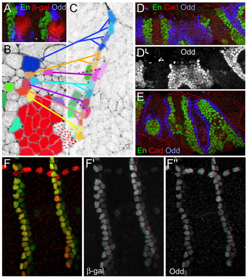
Figure 3. Dynamics of Odd expressing cells.

(A) Odd cells (blue) dorsal and ventral to the tracheal placode, marked by β-gal (red) in stage 9 trachealess-lacZ embryos. (B) and (C) Images from a Cadherin-GFP time-lapse movie (see Movie 1 in supplementary material). Cell shape and disposition allows the identification of the groove and tracheal cells. (C) The epithelium after tracheal invagination. Going back in time shows that groove cells originate from the Odd domain. Tracheal precursors (red), groove cell precursors (blue, yellow, pink, burgundy). Some anterior cells (green) reveal that the groove cells abutting the tracheal placode have been displaced ventrally. Two pink sister cells follow different fates as only one becomes a groove cell.
(D, E) runt3 embryos at stage 12 (D) and 14 (E) display refinement of Odd expression. First, Odd expression (blue; D′) decreases away from the En cells (green); later a single cell wide stripe remains (E).
(F) Odd lacZ shows that β-gal remains visible (green; F′) in cells that have lost Odd. (red; F″)
