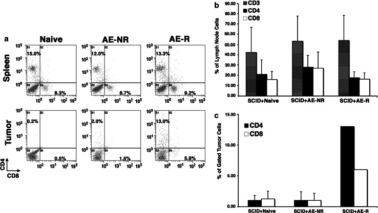
Fig. 2.
Reconstitution of SCID mice by adoptive T cell transfer and homing to tumors. a Spleens, lymph nodes, and tumors from mice of each group that was adoptively transferred (see Fig. 1) were evaluated for the presence of CD4 and CD8 T cells as a proportion of all viable cells. The top panels show staining on representative spleen samples. The bottom panels show staining for T cells in tumors from the same mice in each group. Tumors were harvested at day 15 for the mice reconstituted with naïve T cells and with antigen-experienced T cells that did not lead to rejection in the donors (AE-NR), and at day 18 from the single mouse that developed a tumor from the group reconstituted with antigen-experienced T cells from donors that rejected their tumors (AE-R, FasL-primed). A region was created around the area where lymphocytes scatter light. CD3+, CD4+, and CD8+ cells were analyzed within that population. Light scatter signatures of CD3+ cells in tumors were comparable to those seen in spleens and lymph nodes. CD4 T cells are shown on the Y-axis and CD8 cells in the X-axis. b Graphical representation of means ± SD (measured in three mice from each group) for the percentage of CD3, CD4, and CD8 T cells as a function of all viable cells in reconstituted lymph nodes. These cell frequencies were equivalent to those found in wild-type mice. Similar data were obtained from spleens from four additional mice examined (not shown). c Graphical representation of means ± SD (measured at day 15 in four mice from each control group, and at day 19 in the single mouse from the FasL-T group that developed a tumor) for CD4, and CD8 T cells in tumors

