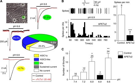
Figure 1.
ASIC3 senses cutaneous acidic pain in rat. (A) Quantification and analysis of pH 6.6-evoked ASIC currents recorded at −80 mV from rat skin DRG neurons using the PcTx1 toxin. Skin DRG neurons in primary culture were identified using fluorescence microscopy after retrograde labelling with DiI (see the image on the top left). The respective percentages of the different current types are highlighted under each current trace and on the graph (data obtained from a total of 43 neurons). (B) Exemplar response of a CM-fibre to pH 6.9 (spikes) with the corresponding time plot of the spike-frequency shown below. The firing of action potential is maintained at pH 6.9, and application of APETx2 10 μM (bar) inhibits the response. The top trace shows the average action potential. Average spike frequency at pH 6.9 and pH 6.9 with APETx2 10 μM (n=9) is represented on the left. (C) Effect of moderate subcutaneous acidification on pain behaviour in rat (determined as the number of flinches of the injected hind paw; see Materials and methods). The injected solution (NaCl 0.9%+20 mM HEPES) was buffered at pH values ranging from 7.4 to 6.6. Condition in which APETx2 10 μM was added to the injected solution is represented in black (*P<0.05 and **P<0.01, significantly different from pH 7.4, Kruskal–Wallis test followed by a Dunn's post hoc test).

