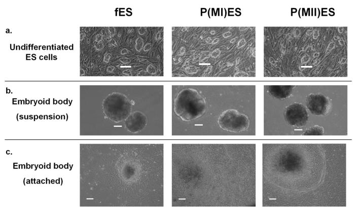
Figure 1.
Undifferentiated and differentiating embryonic stem cells derived from fertilized and parthenogenetic embryos. Fertilized ES cells (fES) and parthenogenetic ES cells [p(MI)ES and p(MII)ES] showed similar morphologies as undifferentiated cells (a), as embryoid bodies in suspension (b) and as attached embryoid bodies (c). Images were captured using a phase contrast microscope. Scale bar = 100 μm.

