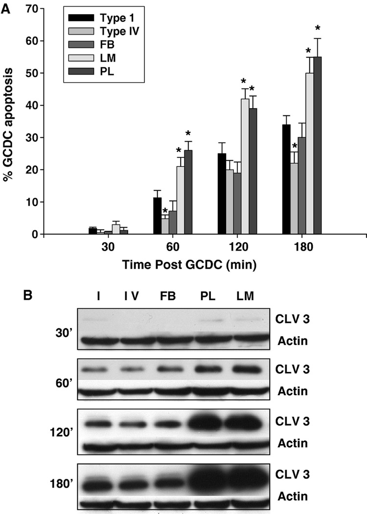
Figure 1. The extracellular matrix modulates hydrophobic bile acid induced rat hepatocyte apoptosis.
Primary rat hepatocytes were plated on Type 1 collagen (I), Type IV collagen (IV), fibronectin (FB), laminin (LM) or polylysine (PL) and the amount of apoptosis after exposure to 50 uM glycochenodeoxycholate (GCDC) determined at the indicated time points. Apoptosis was monitored morphologically using Hoechst staining (A) and biochemically by immunoblotting for the 17/19 kd cleavage fragment of caspase 3 (B). Cleaved 3 immunoblots were re-probed with actin to assure equal protein loading. The results represent the mean and standard deviation of at least 5 separate experiments. The * indicates the value is statistically different than the amount of apoptosis seen in hepatocytes plated on Type 1 collagen at that time point.

