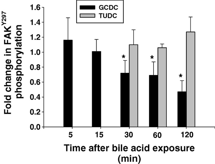
Figure 5. Effect of bile acids on FAK phosphorylation.
Hepatocytes plated on to Type 1 collagen were treated with 50 uM tauroursodeoxycholate (TUDC) or 50 uM glycochenodeoxycholate (GCDC) and the amount of FAKTyr397 phosphorylation at the indicated time points determined by immunoblotting. The results represent the mean and standard deviation of 5 separate experiments. The * indicates the value is statistically different than in control untreated hepatocytes.

