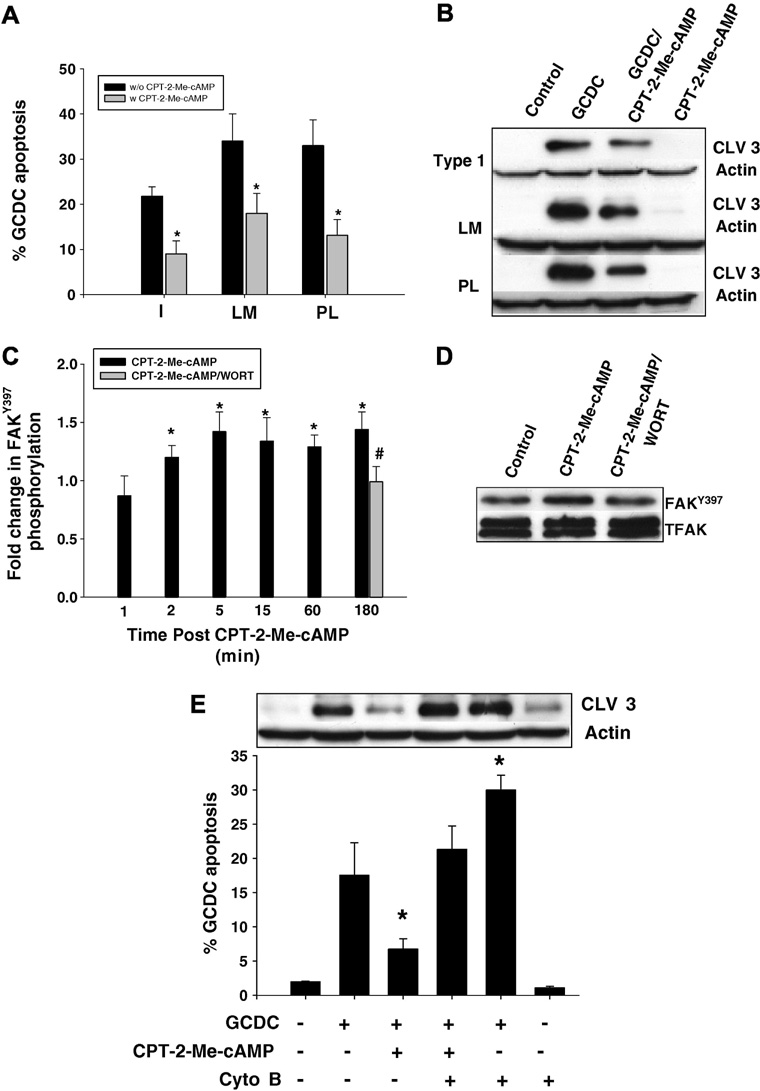
Figure 6. The cytoprotective effect of CPT-2-Me-cAMP is ECM independent but dependent on PI3K mediated FAK phosphorylation.
Hepatocytes were plated on Type 1 collagen (I), laminin (LM) or polylysine (PL). Some cultures were pre-treated with 20 uM CPT-2-Me-cAMP for 20 minutes, and subsequently the amount of apoptosis after 2 hr treatment with 50 uM glycochenodeoxycholate (GCDC) was determined by morphologic evaluation of Hoechst stained cells (A) and by immunoblotting for the 17/19 kD cleavage product of caspase 3 (B). The * indicates the value is statistically different than that seen in the presence of GCDC alone. C. Hepatocytes plated on Type 1 collagen were treated with 20 uM CPT-2-Me-cAMP for the time indicated +/− pretreatment for 15 min with 50 nM wortmannin (WORT) and then the amount of FAKTyr397 determined by immunoblotting. Representative immunblots for the 180 min time point are shown in D. The * indicates the value is statistically different than seen in control cultures and the # indicate the value is different from that seen with CPT-2-Me-cAMP. E. Hepatocytes plated on Type 1 collagen were treated with 1 uM cytochalasin B (Cyto B), 20 uM CPT-2-Me-cAMP or sequentially with Cyto B followed by CPT-2-Me-cAMP and then apoptosis induced by 2 hr treatment with GCDC and quantified by morphologic evaluation of Hoechst stained cells or biochemically by immunoblotting for the 17/19kD cleavage fragment of caspase 3. The * indicated the value is significantly different from that seen in GCDC treated hepatocytes. The results in all the figures represent the mean and standard deviation of 4 experiments.

