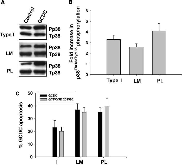
Figure 9. Effect of ECM on bile acid induced phosphorylation of p38 MAPK.
Hepatocytes were plated on Type 1 collagen (I), laminin (LM) or polylysine (PL) and the amount of p38 MAPK phosphorylation determined by immunoblotting 6 hours later. Representative immunoblots are shown in A and B is the quantification (mean and standard deviation) of 3 separate experiments. C. Hepatocyte adhered to I, LM or PL were treated with 50 uM glycochenodeoxycholate (GCDC) for 2 hrs +/− 15 minute pretreatment with 5 uM SB 203580 and the amount of apoptosis determined morphologically by evaluation of Hoechst stained cells. The results are the mean and standard deviation of 3 separate experiments.

