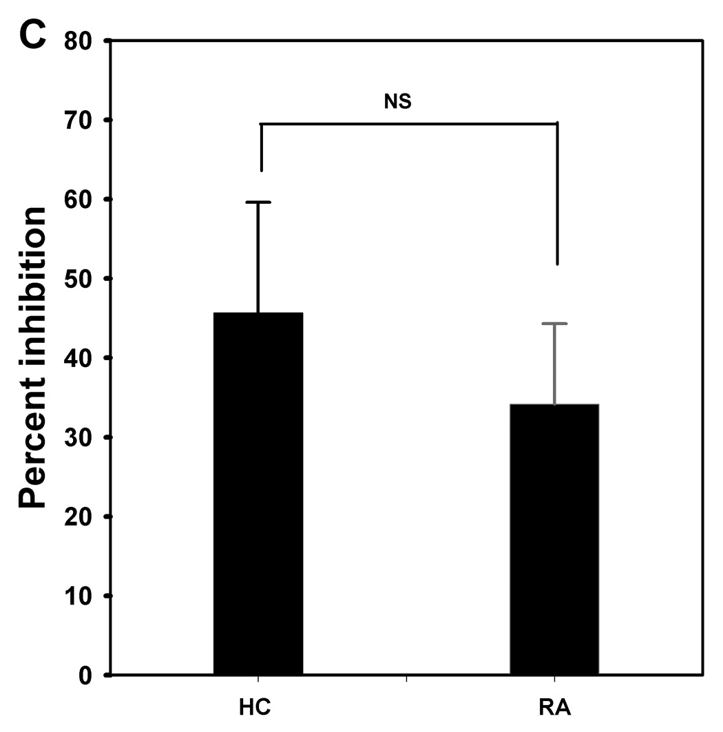
Figure 3. CD4+CD25high T cells isolated from the peripheral blood of patients with RA have suppressive activity.


CD25high PBMC and CD25− PBMC were isolated using a magnetic bead method (as described in Materials and Methods) from peripheral blood of HCs (n=4) and patients with RA (n=6) Representative dot plots of the CD25high PBMC and CD25− PBMC populations are shown (A). Isolated or combined (1:1) CD25− PBMC and CD25high PBMC were polyclonally stimulated with plate-bound anti-CD3 and soluble anti-CD28 antibodies for 5 days, and proliferation was measured for the last 18 hr of incubation using [3H]-thymidine incorporation (B). Results are expressed as mean PSL value ± SEM. In co-cultures, CD25high PBMCs were able to suppress CD25− PBMC proliferation. To assess the suppressive activity of CD25high PBMC, coculture experiments were performed in which CD25−PBMC were cultured in the absence or presence of CD25high PBMC at 1:1 ratio (C), after which the relative difference in proliferative response between the two conditions was calculated. Results are expressed as mean values ± SEM; NS means not significant.
