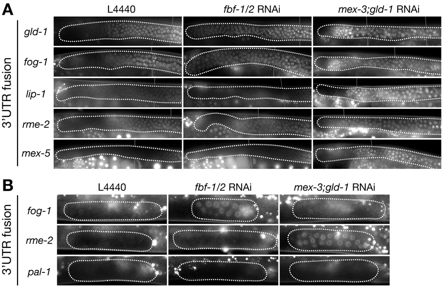
Figure 5. Regulation of 3’UTR fusions in the distal gonad by FBF-1/2, MEX-3 and GLD-1.
A. Fluorescence photomicrographs of distal arms (A, B and C regions in Fig. 1b) from adult hermaphrodites expressing the indicated 3’ UTR fusions, and fed either control bacteria (L4440) or bacteria expressing double-stranded RNA against fbf-1 and fbf-2 or mex-3 and gld-1. See Sup. Table 2 for numbers and additional 3’ UTR fusions examined. Note that lip-1 has low, but non-zero expression throughout the distal region.
B. Photomicrographs of L2 gonadal arms expressing the indicated fusions, and fed either control bacteria (L4440) or bacteria expressing double-stranded RNA against fbf-1 and fbf-2 or mex-3 and gld-1. The RNAi conditions used in these experiments allow the small pool of L2 germline progenitors (~30 cells per arm) to proliferate as in wild-type (~1000 cells per arm by adult stage), indicating that the cells retain mitotic potential and have not yet been transformed. See Sup. Table 3 for numbers and additional 3’UTR fusions examined. Bright foci outside of the gonad are autofluorescent gut granules.

