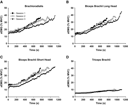
FIG. 3.
Average electromyograph (EMG) at 1% intervals of time to failure for the duration of each practice session for the brachioradialis, long head of biceps brachii, short head of biceps brachii, and triceps brachii (A–D). The r2 values across sessions and muscles for the exponential fits were all >0.87. The rate of increase in biceps brachii short head EMG (C) was significantly lower for session 3 compared with session 1 (P < 0.05).

