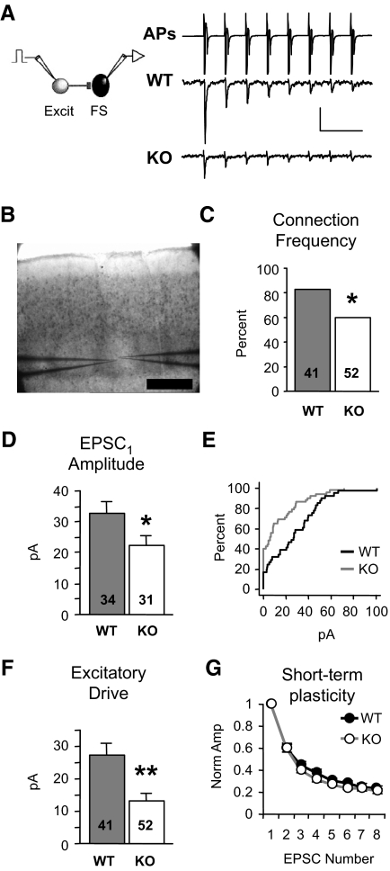
FIG. 1.
Local excitation onto neocortical fast-spiking (FS) inhibitory neurons is dramatically reduced in Fmr1 knockout (KO) mice. A: examples of presynaptic action potentials (APs) being evoked in the excitatory neuron (top, truncated vertically) and resulting unitary excitatory postsynaptic currents (EPSCs) in layer 4 FS neurons (bottom). APs are elicited in voltage clamp and occur due to voltage escape at the site of AP generation. The waveform of the presynaptic neuron represent the currents generated by the APs. EPSCs are averages from single neurons. Scale bars: vertical, 700 and 10 pA for APs and EPSCs, respectively. Horizontal, 50 ms. B: paired recordings were performed inside layer 4 barrels Scale bar: 250 μm. C: the percentage of synaptically connected excitatory and FS inhibitory neuron pairs is reduced in the KO [χ2; number of total cell pairs tested (n) is indicated in each bar; 34/41 and 31/52, connected/total tested]. D: when a connection existed, average amplitude of EPSC1 (1st EPSC in a train) was significantly decreased in the KO. Number of connected cell pairs (n) is indicated in each bar. E: cumulative distribution of amplitudes, including “nonconnected” pairs (0 pA), from wild-type (WT, black) and KO (gray) mice. F: excitatory drive, the average of both connected and nonconnected pairs, is reduced by 51%. Number of total cell pairs tested (n) is indicated in each bar. G: no change in short-term plasticity of EPSCs was observed (n = 22,16 connected pairs; quantified by an STP index as described in methods). Each EPSC in the train is normalized to EPSC1. *P < 0.03, **P < 0.005. Sample numbers as described in the preceding text apply to similar graphs in subsequent figures. See methods for statistics used.

