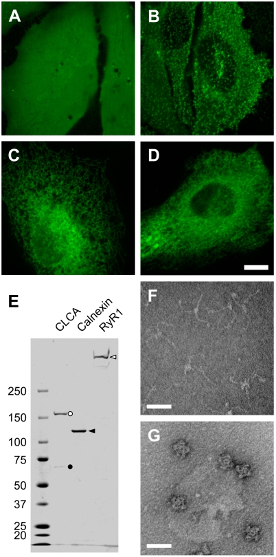
Figure 2. Localization of mfGFP fusion proteins in living cells and isolation of mfGFP-fusion protein complexes.
(A–D) HeLa cells were transfected with expression vectors of either mfGFP alone (A) or mfGFP fused with clathrin light chain A (CLCA) (B), calnexin (C), and type 1 ryanodine receptor (RyR1) (D). Whereas mfGFP alone distributed throughout the cells, fusion proteins were localized at the expected site. Scale bars, 10 µm. (E) mfGFP-fusion protein complexes were isolated by streptavidin column chromatography using SBP-tag. The isolated fractions were processed on an SDS-polyacrylamide gel, and stained with CBB. CLCA (black circle) co-purified clathrin heavy chain (CHC) (white circle). Calnexin and RyR1 were isolated as a single band. (F and G) Negative staining EM observation of isolated CLCA and RyR1 fractions. CLCA exhibited three-armed pinwheel morphology of triskelion (F), whereas RyR1 exhibited characteristic quarterfoil appearance (G). Scale bar, 50 nm.

