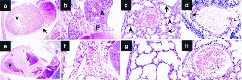
Figure 1.
(a–d) Histological abnormalities in c-Abl−/− p53−/− mice. (e–h) Wild-type newborn sibling control. (a) Section of the heart. Hemorrhage in the pericardial sac (arrow). There is no blood in the left ventricle (V). (e) Both ventricles (V) in the wild-type sibling contain blood. (b) Right lung. Anterior lobe (A) is not expanded. Posterior lobe (P) is minimally expanded. Compare with f. (c) Higher power of the right posterior lobe shows thick walls of the alveolar sacs (arrows) with bulging capillaries (arrowheads). Compare with wild-type sibling shown in g. (d) Arterial wall in the right posterior lung lobe. Note thickness of the media (arrow). Compare with h, which shows the wild-type sibling

