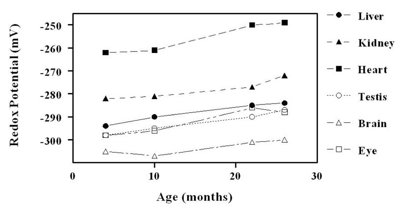
Figure 5.
Glutathione redox potentials in mitochondria from liver, kidney, heart, testis, brain and eye. Redox potentials become significantly (P< 0.01) more pro-oxidizing in the old (26 months) compared to young (4 months) mice in all the tissues, except brain (adapted from [26]). The standard deviation was less than 3–5% (error bars are omitted for the purpose of clarity).

