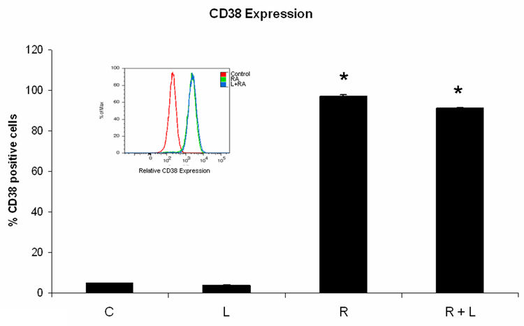
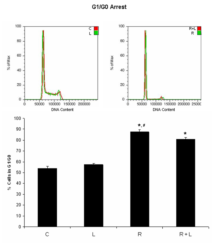
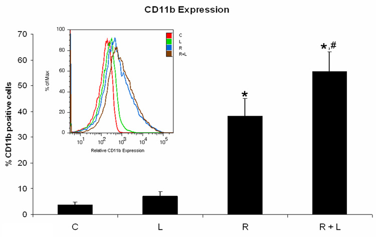
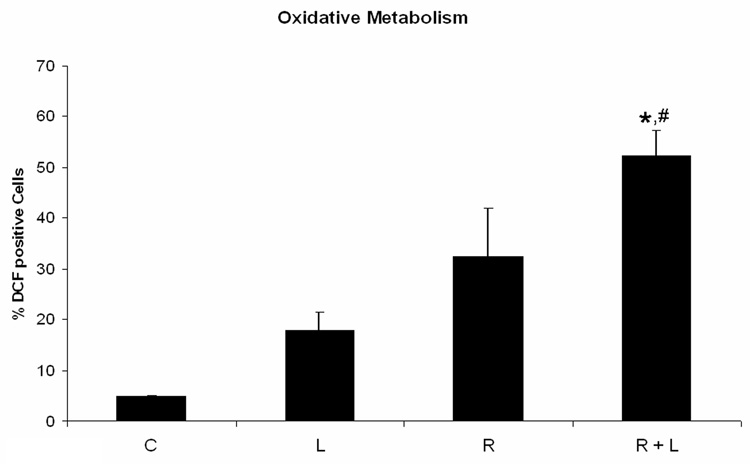
Figure 3. HL-60 cells treated with integrin αMβ2 ligand during myeloid differentiation have enhanced features of myeloid differentiation.
HL-60 cells were treated or not with RA plus or minus integrin ligand for 24 h, 48 h, and 72 h, respectively. CD38 expression at 24 h (A) and G0 arrest at 72 h (B) were decreased in cells treated with RA plus integrin ligand when compared to cells treated with RA alone. CD11b (C) expression at 48 h and the capability of oxidative metabolism at 48 h (D) were increased in cells treated with RA plus integrin ligand when compared to cells treated with RA alone. Shown are means with error bars representing standard errors of means from 3 independent experiments. The insert shows CD38 histograms (panel A), DNA histograms (panel B), CD11b histograms (panel C) for a typical experiment. (*) indicates treatment groups that were significantly different from control and (#) indicates RA plus AG1296 treated groups that were significantly different from cells receiving RA only. C, control; L, 5 µM integrin αMβ2 ligand; R, 2 µM retinoic acid; R + L, 5 µM integrin αMβ2 ligand plus 2 µM retinoic acid.


