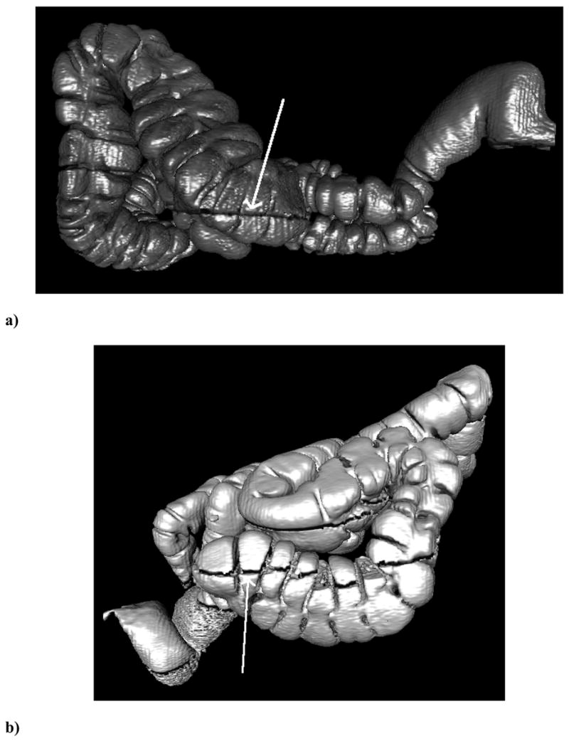
Figure 2.

Lateral surface-rendered three-dimensional reconstructions of the colon from CTC showing representative fluid levels and their automated measurements. The arrows indicate the air/fluid boundary. The CT scanner table (not shown) is at the bottom of the image so that the fluid is dependent. (a) The ascending colon of a 69 year old male on prone CTCs showing low surface area obscured by fluid with a fluid score of 1.2%. (b) The ascending colon of a 54 year old male on supine CTC showing a high surface area obscured by fluid with a fluid score of 83.1%.
