
Figure 3.
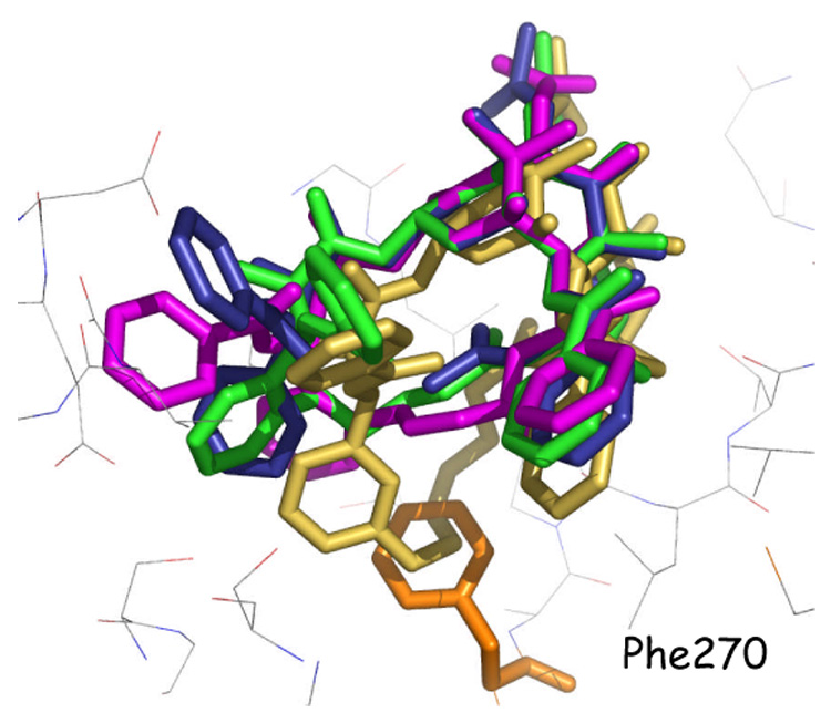
T-Conformation of PTX (blue) bound to β-tubulin. The T-conformers for the analogs E-15c (green) and Z-15g (medium brown) and E-18f (magenta) have been derived from NAMFIS analyses. The baccatin cores for PTX and the three analogs are superimposed. The meta-Z-bridged 15g (light brown) is pushed out of the taxane binding site because phenylalanine 270 of tubulin (orange) is in steric conflict with the extended bridge preventing effective binding to the protein. 15c and 18f avoid the Phe272 steric-clash and fit nicely into the PTX binding site.