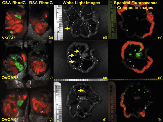
Figure 3.

(a) Side‐by‐side spectral fluorescence composite image of the exposed abdomens of SKOV3 tumor‐bearing mice treated with galactosyl serum albumin (GSA)–rhodamine green (RhodG) (left) and bovine serum albumin (BSA)–RhodG (right) demonstrates brighter tumor‐associated fluorescence with GSA–RhodG than with BSA–RhodG. (b,c) Similar results were observed for OVCAR5 and OVCAR8 tumor‐bearing mice, respectively. (d) White‐light photo of a loop of bowel and mesentery from a SKOV3 tumor‐bearing mouse treated with GSA–RhodG identifies only one tumor nodule (yellow arrow). (e,f) Similar results were observed for OVCAR5 and OVCAR8 tumor‐bearing mice, respectively. (g) Spectral fluorescence composite image of the same tissue shown in 3‐D now reveals multiple tumor nodules (green), indicating that targeted optical imaging can identify more disease than the unaided eye. (h,i) Similar results were observed for OVCAR5 and OVCAR8 tumor‐bearing mice, respectively. (Note: Red = autofluorescence of the gastrointestinal tract.)
