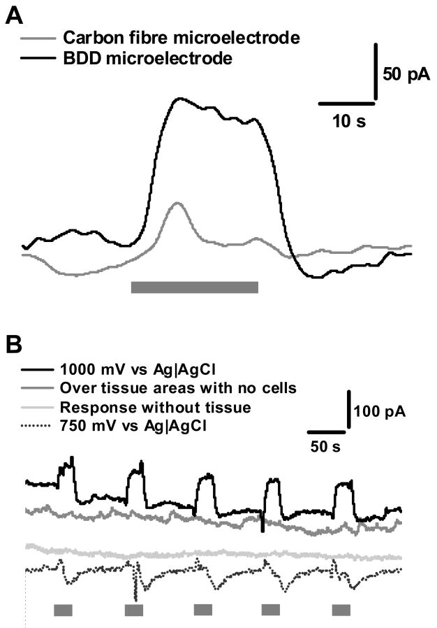
Figure 2.
Experimental controls to show NO detection from myenteric plexus. In all experiments, the grey bar indicates the period during a 20 s superfusion with 1 μM nicotine. This stimulation is provided for all experiments. (A) shows a comparison between a BDD and carbon fibre microelectrode, where both electrodes are held at 1 V. (B) shows the experimental controls, were the electrode is held at a potential to measure NO, when induced using a nicotine stimulation. The controls all show a lack of positive current as would be expected if NO were not present. There are small to no responses when measurements are made without the presence of tissue or in areas outside the ganglionic regions indicating that the majority of release is from NOS containing neurons and terminals from the MP, rather than synaptic terminals within the longitudinal muscle. The trace at 750 mV indicated no interference from catecholamines as no positive deflections in the current were observed.

