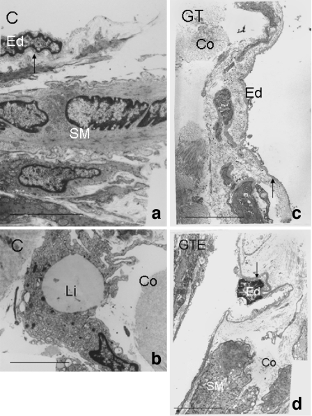
Fig. 4.
Ultrastructure of cavernous tissue of rats from control (C) (a, b), green tea (GT) (c) and green tea extract (GTE) (d) animals. Endothelial cells (Ed) were separated by basal lamina (arrow) from the surrounding smooth muscle fibres (SM). Profuse collagen fibres (Co) were observed in all experimental groups studied. Note the increased average thickness of the basal lamina (arrow) in the corpus cavernosum of rats ingesting GT or GTE (c and d, respectively). In C animals there were frequent lipid droplet rich cells (Li) in erectile tissue, localised close to the vascular space. Bar 5 μm

