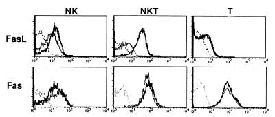
Figure 4.
Expression of FasL and Fas on hepatic MNC subpopulations with or without Con A injection. Wild-type B6 mice were i.v. injected with 15 mg/kg Con A or PBS only. Hepatic MNC were isolated 2 h later, and then stained with biotinylated anti-FasL mAb or anti-Fas mAb followed by FITC-conjugated anti-mouse NK1.1 mAb, Cy-chrome-conjugated anti-CD3 mAb, and PE-conjugated streptavidin. Expression of FasL or Fas was analyzed on electronically gated NK1.1+CD3− (NK), NK1.1+CD3+ (NKT), or NK1.1−CD3+ (T) cells. Bold lines indicate the staining of Con A-injected hepatic MNCs, thin lines indicate the staining of PBS-injected hepatic MNCs, and broken lines indicate the background staining with isotype-matched control IgG. Similar results were obtained in three independent experiments.

