
Figure 1. GSK3 inhibitors and siRNA result in glioma cell death and reduced tumorigenicity.
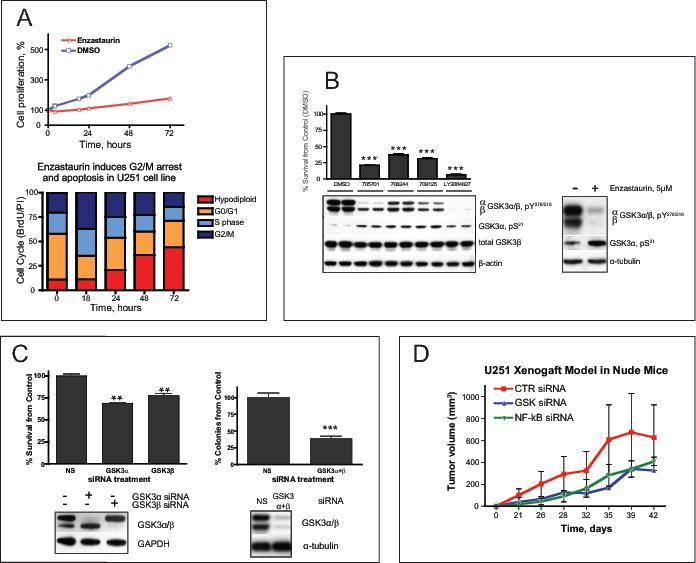
(A) Cytotoxicity of enzastaurin (5 μM) in U251 glioma cell line as determined by a cell-counting assay at different time points as indicated. Bar graph shows results of the cell cycle analysis of U251 cells exposed to enzastaurin for the indicated time, then labeled with10 μM BrdU for 2 hours The drug induces G2/M arrest at 18 hours and subsequent apoptosis. (B) Changes in phosphorylation of GSK3 at Y276/216 and S21 of GSK3α caused by different GSK3 inhibitors correlate with survival of U251. Cells were treated with 0.5 μM of the indicated drugs for 48 hours Cell survival was assessed by cell counting (upper panel, graph). Y216/279 activating phosphorylation and S21 inhibitory phosphorylation of GSK3α/β were measured by Western Blot (lower panel). Here and below the asterisks designate p-values (t-test) as follows: *, p<0.05 ; ** , p<0.005 and ***, p<0.00, the error bars represent standard deviation. (C) GSK3 siRNA inhibits proliferation of glioma cells in vitro and in vivo (upper left corner). GSK3-specific siRNAs were transfected into U251 cells. Cells were harvested and counted 48 hours after transfection. Western blot shows efficiency of protein silencing by siRNA. In vitro clonogenicity assay demonstrated reduced number of colonies derived from U251 cells treated with GSK3 siRNA compared to non silencing (NS) siRNA. (D) Subcutaneous xenograft model in nude mice. After treatment with control, GSK3, or NF-κB siRNAs, 0.5 million U251 cells were injected subcutaneously into the thighs of nude mice. Each group consisted of 6 animals. Tumors were measured twice a week. The results are presented as tumor volume.