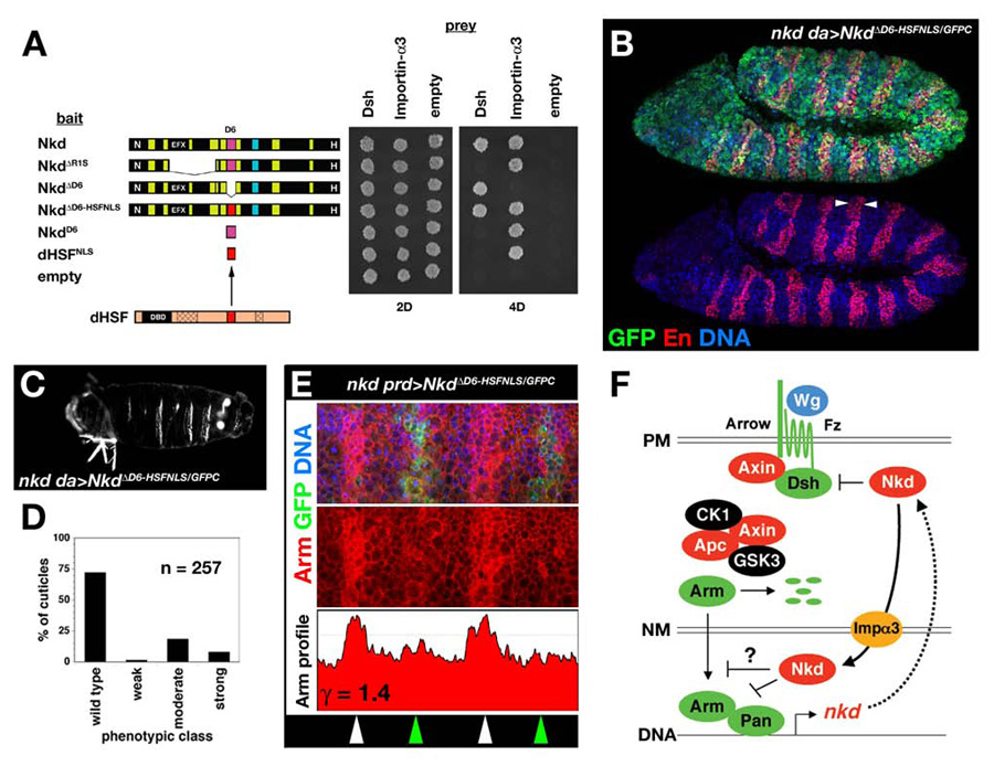
Fig. 7.

Restoration of Importin-α3-association by the dHSF-NLS does not restore Nkd function. (A) Y2H showing that replacement of the D6-NLS (magenta) with the dHSF-NLS (red) restored the Nkd/Importin-α3 interaction but did not affect Nkd/Dsh interactions. (B) Stage 11 nkd da>NkdΔD6-HSFNLS/GFPC stained for GFP (green), En (red), and DNA (blue) with wide En stripes (arrowheads). (C) Representative nkd da>NkdΔD6-HSFNLS/GFPC cuticle, with 3 complete denticle belts indicative of moderate-class nkd cuticle. (D) Distribution of nkd cuticle phenotypes for NkdΔD6-HSFNLS/GFPC rescue cross (n=number of cuticles scored). (E) Stage 10 nkd prd>NkdΔD6-HSFNLS/GFPC embryo stained for GFP (green), Arm (red), and DNA (blue) in top panel, with Arm-only channel and Arm greyscale intensity profile (ordinate axis 0–200 pixels) in middle and lower panels, similar to Fig. 1C. (F) Model for fly Nkd function. Wg signaling at the plasma membrane (PM) leads to Arm/Pan-dependent transcription of target genes, including nkd (see Introduction for details). Nkd targets Dsh in the cytoplasm and/or plasma membrane, and binds Importin-α3 to traverse the nuclear membrane (NM). In the nucleus, whether Nkd inhibits nucleocytoplasmic transport of Arm or another molecule, or whether Nkd directly regulates target gene transcription, remains unknown.
