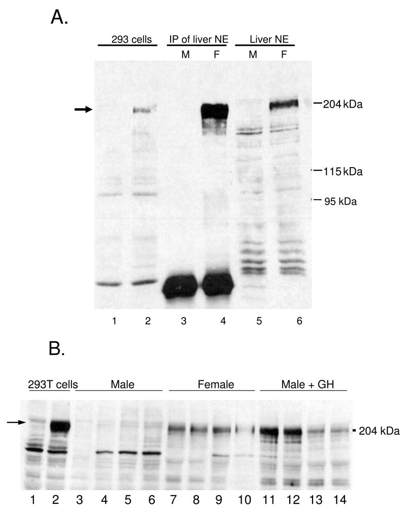
Figure 2. Western blot analysis of liver Cutl2 protein.
Nuclear extracts prepared from pooled mouse livers (panel A) or from individual rat livers (panel B) were resolved on a 6% SDS polyacrylamide gel and subjected to Western blot analysis with anti-Cutl2 antibody 356 in comparison to cDNA-expressed mouse Cutl2. Panel A: whole cell extracts (40 μg) from untransfected 293T cells (lane 1) or from 293T cells transfected with Cutl2 cDNA (lane 2); Cutl2 protein immunoprecipitated with anti-Cutl2 antibody 356 from liver nuclear extract (1 mg) prepared from pools of adult male (lane 3) and female (lane 4) mouse liver (n=8 livers/group); and adult male (lane 5) and female (lane 6) mouse liver nuclear extract (40 μg/lane). Panel B, portion of Western blot showing, in lanes 1–2: cell extracts (40 μg) from untransfected or Cutl2 cDNA-transfected 293T cells, respectively; lanes 3–14: liver nuclear extracts prepared from individual adult male (lanes 3–6) and female (lanes 7–10) rats and from adult male rats given a continuous GH infusion for 7 d (lanes 11–14). Authentic, cDNA-expressed Cutl2 protein (lane 2, panels A and B; arrow) runs close to the 204 kDa protein marker shown on the right.

