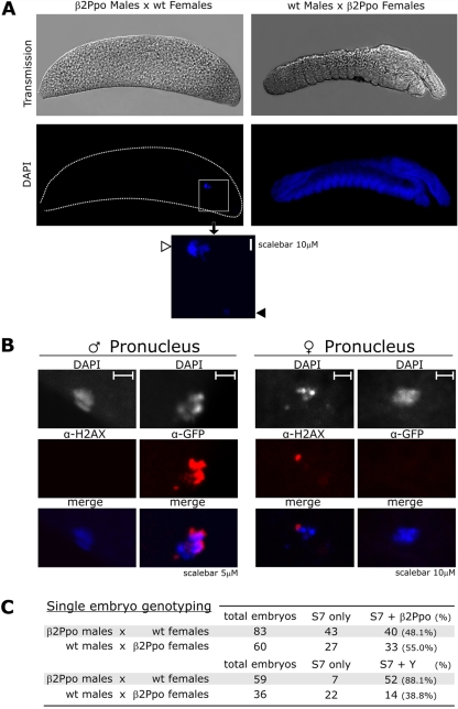
Figure 3. Morphological and genotype analysis of developmentally arrested embryos.
(A) Embryos originating from crosses between β2Ppo males with WT females (left) compared to crosses between WT males and β2Ppo females (right) were analyzed by fluorescence microscopy 24 hours after oviposititon. The figure shows transmission (upper panels) and fluorescence of DAPI staining DNA (lower panels) of embryos oriented with the posterior end to the left and ventral side up. The inset shows a magnified view of the small and large DAPI stained bodies found in the embryos marked with a black and a white arrow respectively. (B) Immunostaining of freshly hatched embryos using mouse anti GFP (α-GFP) or mouse anti γ-H2AX (α-H2AX) primary in combination with anti-mouse IgG Alexa-532 conjugated secondary antibodies. DAPI stained bodies identified as male pronucleus and female pronucleus are shown at 5 and 10 µM scale respectively. (C) Molecular genotyping of embryos using multiplex PCR. Embryos originating from crosses of β2Ppo males with WT females and WT males with β2Ppo females were collected at 24 hrs post deposition and their DNA was examined using nested PCR analysis to amplify Y chromosome or transgene specific sequences as well as the ribosomal gene S7 as a control. The values show the frequency of the genotypes in all embryos that tested positive for the presence of S7.

