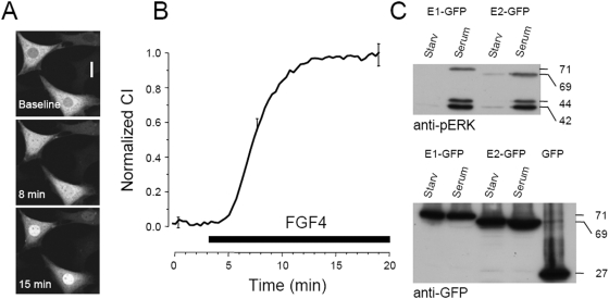
Figure 1. ERK1-GFP translocates in the nucleus of NIH-3T3 cells after stimulation.
A) Cells transfected with ERK1-GFP in control conditions and after treatment with 80 ng/ml of FGF4 (calibration bar 20 µm). B) Time course of the normalized Concentration Index of cells stimulated with FGF4 (n = 18). The vertical bars are the standard error of the mean at the given point and are representative of the experimental variability of the entire data set. C) ERK-GFP fusion proteins are phosphorylated following serum stimulation, as demonstrated by western blot with a phospho-specific ERK antibody (upper panel). The fusion proteins have the correct molecular weight also when assayed with an anti-GFP antibody.

