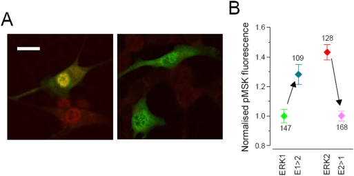
Figure 9. Effects of the overexpression of N-terminus mutations of ERK1/2 on the nuclear target MSK.
A) Representative fields of fibroblast treated for 24 hr with 10% serum. In this example, cells have been transfected with ERK2-GFP and were stained with the pMSK specific antibody (red). The left panel show that the treatment caused nuclear translocation of ERK2-GFP and activation of MSK, which is well visible both in the transfected cell (the nucleus is yellow) and in nearby non-transfected cells. The inhibitor U0126 (25 µM) was administrated 6 hr before fixation. In these conditions ERK did not accumulate in the nucleus and there was not detectable phosphorylation of MSK, indicating that, MSK requires ERK activity to be phosphorylated after serum treatment. Bar 20 µm. B) Intensity of the pMSK signal measured in cells transfected with ERK1, ERK2, E1Δ39 (indicated as E1>E2) and Δ39E2 (indicated as E2>E1). The experiment was repeated in triplicate and the fluorescence of each cell has been normalised to the mean fluorescence of the ERK1 group to allow the comparison of the different experiments. Number of cells indicated nearby each symbol. U0126 suppressed almost completely the pMSK signal (mean = 0.48±0.04, n = 76; not shown). Since cell fluorescence was not normally distributed, significativity was assessed with the Kolmogorov-Smirnoff test. ERK1≠ERK2 (p≤0.0005); E1>E2≠E2>E1 (p≤0.0005). E1 and E2>E1 are not significantly different (p≤0.2). E1>E2 is larger that ERK1 (p≤0.0005) but it is also slightly but significantly smaller than ERK2 (p≤0.01).

