Figure 2.
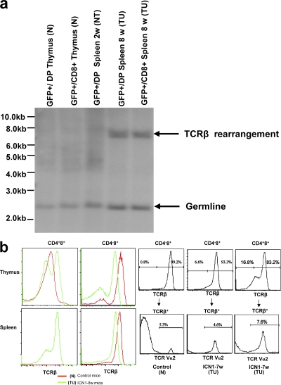
TCR expression by malignant and nonmalignant cells. (a) TCR-β rearrangement in various cell types. Analysis of TCR-β rearrangement by Southern blot analysis in GFP+CD4+8+(GFP+/DP) and GFP+CD4−8+(GFP+/CD8+) thymocytes derived from retroviral GFP-empty vector–transplanted mice, as well as GFP+CD4+8+(GFP+/DP) and GFP+CD4−8+(GFP+/CD8+) splenocytes derived from retroviral GFP-ICN1–transplanted mice at 2 and 8 wk after BMT. Molecular markers are shown on the left. (b) TCR-β and TCRV-α2 expression by various lymphocyte subsets. (left) Staining with the pan TCR-β antibody H57. (right) Staining with Vα2 TCR antibody.

