Figure 2.
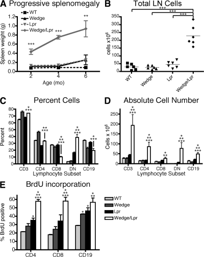
Lymphocyte expansion in CD45 wedge/lpr mice is accompanied by increased cell proliferation. (A) Splenic weight of 2-, 4-, and 6-mo-old mice. Mean of three to six mice per time point ± SEM. ANOVA: *, P < 0.05; **, P < 0.005; ***, P < 0.0005. (B) Total number of cells pooled from inguinal, axillary, brachial, cervical, and mesenteric lymph nodes of 2-mo-old mice. Unpaired Student's t test: ***, P < 0.0005. (C) Lymphocyte subset analysis of lymph nodes from 2-mo-old mice. CD3+ cells are separated into CD4+, CD8+, and DN (CD3+, B220+, CD4−/CD8− [DN]) cells. (D) Mean absolute number of T cell subsets and B cells in lymph nodes from 2-mo-old mice. Error bars represent SEM with two to four mice per group from three independent experiments. (E) BrdU incorporation is increased in CD4 and CD8 T cells and B cells from wedge/lpr mice. 7–8-wk-old mice were given BrdU in their drinking water for 10 d. Shown is the mean percentage of cells positive for intracellular BrdU from four mice per genotype ± SEM. Data are representative of three independent experiments. (C, D, and E) Unpaired Student's t test, P < 0.05 compared with WT, *; wedge, **; and lpr, ***.

