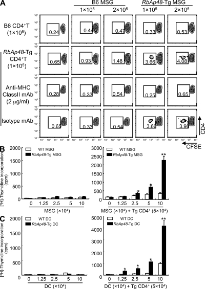
Figure 4.
CD4+ T cells can proliferate to epithelial cells from RbAp48-Tg mice. (A) CFSE-labeled purified CD4+ T cells (105) of WT and RbAp48-Tg mice were co-cultured with MSG cells (1 and 2 × 10⁵) from the mice for 72 h. Cell proliferation was estimated by the dilution of CFSE. 2 μg/ml anti–MHC class II mAb or isotype control antibody was added in the culture. All results are representative of three to five mice at 28 wk of age, or three-independent experiments. (B) CD4+ T cells (5 × 10⁴) of cLNs from RbAp48-Tg mice were co-cultured with irradiated MSG cells (0–10 × 104) from WT and RbAp48-Tg mice for 72 h. (C) CD4+ T cells (5 × 10⁴) of cLNs from RbAp48-Tg mice were co-cultured with irradiated DCs (0–10 × 104) from WT and RbAp48-Tg mice for 72 h. Proliferative T cell response was evaluated by [3H]thymidine incorporation during the last 12 h of the culture. Results are representative of means ± SE of triplicates in two independent experiments. *, P < 0.05; **, P < 0.005; WT versus RbAp48-Tg MSG cells or DCs.

