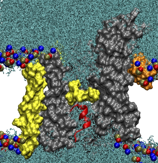
Figure 5.
Modification of the plug's interactions with water. SecYEβ is shown as a molecular surface, colored as in Fig. 1, cut through the middle to display the channel. Lipid head groups are indicated as blue, red, and brown spheres. Water molecules, shown in light blue, fill the channel up to the pore ring, in yellow, from both top and bottom. The water molecules clearly overlap the plug, shown in red.

