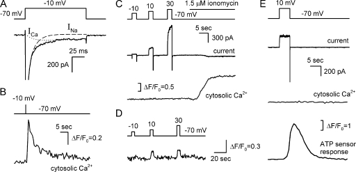
Figure 3.
ATP release on electrical stimulation is not accompanied by a change in intracellular Ca2+. (A and B) Representative simultaneous recordings of ionic currents and fluorescence in a type III cell loaded with 4 μM Fluo-4 (n = 6). The depolarization of the taste cell to −10 mV for 100 ms (A and B, top panels) elicited the VG Ca2+ current (A, bottom panel, dotted line) that produced the Ca2+ transient monitored as an increase in Fluo-4 fluorescence (B, bottom panel). A relative change in fluorescence intensity is expressed as ΔF/F0, where F0 is a value of Fluo-4 emission measured just before cell stimulation. (C and D) Representative concurrent recordings of ionic currents (middle panel) and Fluo-4 fluorescence (bottom panel) in a type II cell (n = 21). Unlike ionomycin, 2-s depolarization (top panel) did not affect intracellular Ca2+ in 17 of 21 type II cells (C). In 4 out of 21 cells, depolarization induced a small increase in Fluo-4 fluorescence (D). (E) Representative concurrent recordings of a membrane current and intracellular Ca2+ in a type II cell and an ATP sensor response (n = 9). The 5-s depolarization (top panel) of the taste cell produced no change in cytosolic Ca2+ (the second trace from the bottom) but triggered ATP release, thereby stimulating the ATP sensor (bottom panel). The recording conditions were as in Fig. 1.

