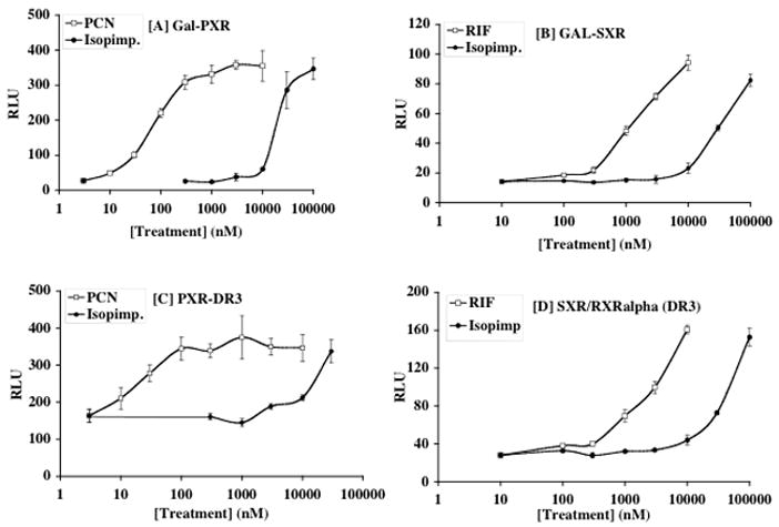
Figure 6.

CV-1 cells were cotransfected with Gal-L-PXR (A) or Gal-L-SXR (B) together with MH100-tk-luc, CMX-β-gal. In panels (C) and (D), cells were transfected with full length PXR or SXR, together with hRXRalpha, (DR3)3-tk-luc and CMX-β-gal. Transfected cells were incubated with media containing indicated amount of ligand or solvent control for 18–24 h before subjected to luciferase and β-gal assay. Transcriptional activation was assessed by the relative luciferase activity normalized by β-gal activity. Figures represent means ± SD.
