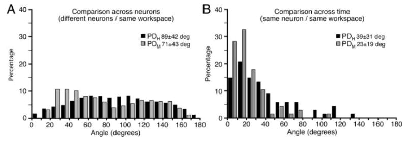
FIG. 6.

Distribution of angles measured between pairs of PDH (black) or PDM (gray) vectors. A: angles between the PDs of all possible combinations of neurons measured within a single workspace. There were somewhat more small angles (similar PDs) within muscle space than hand space, perhaps reflecting clusters of functionally similar neurons. B: angles between repeated estimates of the PD of a given neuron over time within the same workspace. PDM vectors were significantly more stable over time than were PDH vectors.
