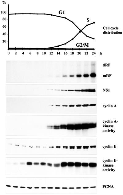
Figure 1.
Time course analysis of MVM DNA replication in synchronized A9 cells. Suspension cultures were synchronized in G0/G1 by serum starvation and infected with MVM (MOI = 10 pfu/cell). At 12 h p.i., cells were released from the G1 block by addition of FCS (20% final concentration). Culture samples were taken at 2-h intervals and monitored for cell cycle distribution, production of MVM DNA replication intermediates, and viral and cellular protein expression. Cyclin A- and E-associated kinase activities were determined as described in Materials and Methods. mRF, monomer replicative form DNA; dRF, dimer replicative form DNA.

