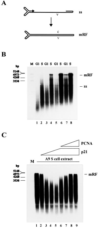
Figure 2.
Conversion of MVM ss DNA into RF DNA in extracts from A9 cells synchronized in G1 and S phase. (A) Schematic representation of the conversion reaction. (B) MVM ss DNA (20 ng) was incubated in G1 or S cell extracts corresponding to 40 μg (lanes 1 and 2), 60 μg (lanes 3 and 4), 80 μg (lanes 5 and 6), or 100 μg (lanes 7 and 8) of cellular proteins. (C) MVM ss DNA (20 ng) was incubated in S cell extract alone (lane 1) or supplemented with 0.4 μg (lane 2), 0.8 μg (lane 3), 1.2 μg (lane 4), or 1.6 μg (lanes 5 to 9) of recombinant p21WAF1/CIP1 and additionally 0.4 μg (lane 6), 0.8 μg (lane 7), 1.2 μg (lane 8), or 1.6 μg (lane 9) of recombinant PCNA. Replication products were analyzed by neutral agarose gel electrophoresis (0.8%). ss, single-stranded virion DNA; mRF, monomer replicative form DNA; v, viral strand; c, complementary strand; M, DNA size markers in bp.

