
Figure 3.
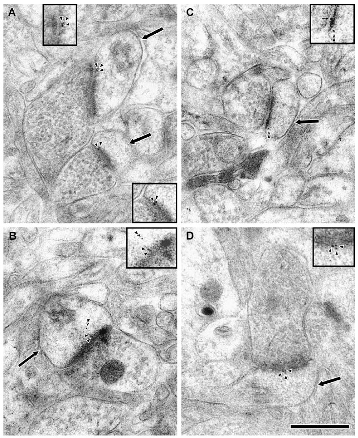
Electron micrographs of axospinous synapses in freeze-substituted material labeled for PP1α and PP1γ1 by post-embedding immunogold. Dendritic spines (arrows) receive asymmetric synapses from axon terminals. Immunoreactivity was revealed by 10 nm gold particles (arrowheads), and the insets show a magnified view of the gold particles. Gold particles labeling for PP1α (A, B) and PP1γ1 (D) were seen in the PSD. PP1α immunoreactivity was also commonly identified in deeper subjacent intraspinous domains (B and C, double arrowhead). PP1α labeling was also observed in the synaptic cleft (C, small arrow). The scale bar is 500 nm.