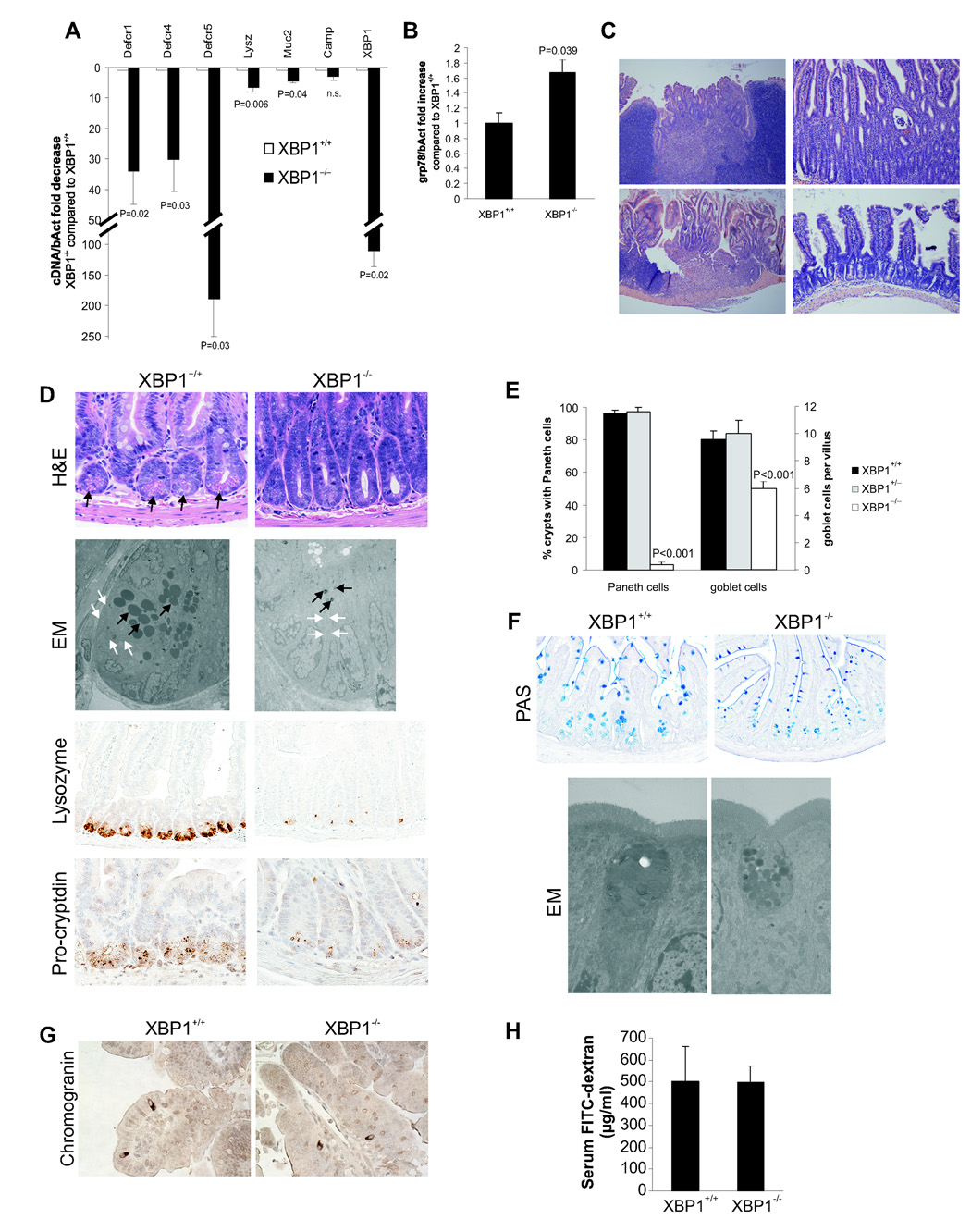
Figure 1. Spontaneous enteritis and Paneth cell loss in XBP1−/− mice.
A. Small intestinal mucosal scrapings (n = 8 per group) from Xbp1-deleted (“XBP1−/−”) and Xbp1-sufficient (“XBP1+/+”) mouse intestinal epithelium were analyzed for cryptdin-1 (Defcr1), cryptdin-4 (Defcr4), cryptdin-5 (Defcr5), lysozyme (Lysz), mucin-2 (Muc2), cathelicidin (Camp1), and XBP1 (primers binding in the floxed region) mRNA expression. Data are expressed as fold decrease in XBP1−/− compared to XBP1+/+ specimens, normalized to β-actin (Student’s t test). B. Fold increase in grp78 mRNA expression in XBP1−/− compared to XBP1+/+ epithelium, normalized to β-actin (n = 3 per group, Student’s t test). C. Spontaneous enteritis in XBP1−/− mice (upper panels and lower left panel), and normal histology of XBP1+/+ mice (lower right panel). Upper left, cryptitis with villous shortening, crypt regeneration and architectural distortion; upper right, neutrophilic crypt abscesses; lower left, duodenitis with surface ulceration and granulation tissue. D. Paneth cells with typical eosinophilic granules on H&E stained sections at the base of crypts in XBP1+/+, but not XBP1−/− epithelium. Electron microscopy (EM) with only rudimentary electron-dense granules and a contracted ER in XBP1−/− basal crypt epithelial cells, normal configuration in XBP+/+ mice. Immunohistochemistry (IHC) for the granule proteins lysozyme and pro-cryptdin in XBP1+/+ and XBP1−/− epithelia. E. Enumeration of Paneth cells and goblet cells in small intestines (n = 5 per group, Student’s t test). F. Goblet cell staining by PAS in XBP1+/+ and XBP1−/− epithelia. EM exhibited smaller cytoplasmic mucin droplets and a contracted ER in XBP1−/− goblet cells. No structural abnormalities were found in neighboring absorptive epithelia in XBP1−/− mice. G. The marker for enteroendocrine cells, chromogranin, was detected by IHC in small intestines of XBP1+/+ and XBP1−/− mice. H. XBP1+/+ and XBP1−/− mice were orally administered FITC-dextran, and FITC-dextran serum levels assayed 4h later.

