



Synopsis
Nearly half of all patients with heart failure have preserved ejection fraction (HFpEF). HFpEF patients tend to be older, female, and hypertensive, and characteristically display increased ventricular and arterial stiffening. In this review, we discuss the pathophysiology of abnormal ventriculoarterial stiffening and how the latter affects ventricular function, cardiovascular hemodynamics, reserve capacity, and symptoms. We conclude by exploring how novel treatment strategies targeting abnormal ventricular-arterial interaction might prove useful in the treatment of patients with HFpEF.
Introduction
The cardiovascular system is designed to provide ample pressure and flow to the body at rest and over broad ranges of stress. Because blood flow is pulsatile, changes in cardiac output are accompanied by alterations in the arterial pulse wave amplitude and pressure. In health, the heart-artery system is compliant to prevent wide swings in pressure that otherwise can lead to vascular and end-organ damage. For the vasculature, this compliance is largely contained within the proximal conduit vessels, and the stiffness (elastance) achieved during contraction by the left ventricle is closely matched or coupled to this arterial elastance to optimize mechanical efficiency and maintain a normal ejection fraction. Perhaps more important than the coupling ratio of ventricular and vascular stiffness are their absolute values. Indeed, maintaining low ventricular and arterial elastances in the normal human allows a dynamic range of volume transfer to be achieved during ejection with minimal change in pressure.
With advancing age, both ventricular and arterial stiffness increase, changes that are further amplified by comorbidities such as hypertension, diabetes, and kidney disease. This stiffening greatly augments systolic and pulse pressure swings during ejection, increasing arterial pressure decay during diastole and ventricular afterload. When incident (ejected) pressure waves encounter zones of impedance mismatch (e.g. arterial bifurcations), part of the wave is reflected backwards, interfering with flow but augmenting pressure. Pressure and flow wave transmission is enhanced in stiff blood vessels, thus the principal reflected wave returns to the heart in late systole, exacerbating systolic load. This increase in net and late-systolic afterload importantly alters ventricular systolic and diastolic function, increasing the amount of hydraulic work (and myocardial oxygen consumption) needed to provide the body with a given amount of blood flow. This in turn leads to impaired cardiovascular reserve function, labile systemic blood pressures, diminished coronary flow reserve, and increases in diastolic filling pressures. Greater pulse perfusion in a stiff arterial system is a risk factor for vascular disease, and may compromise endothelial-dependent vasorelaxation. One can consider this adverse interaction between stiff heart and arteries as a form of coupling disease which ultimately limits the ability of the integrated cardiovascular system to respond to stress.
Ventricular-arterial stiffening is common in patients with heart failure and apparent preservation of systolic function. Such patients are typically older, female, hypertensive, and display a high prevalence of diabetes, obesity, and renal dysfunction. They often develop marked systolic hypertension under conditions of stress, and both their arterial and ventricular systolic pressures are very sensitive to blood volume status. While abnormal diastolic function is thought to contribute to failure symptoms by increasing congestion, it cannot explain the observed increases in systemic pressures nor does it fully underlie limitations of cardiac reserve. Recent animal and human data has refocused attention on the impact of arterial afterload on ventricular diastolic relaxation and compliance. Here, we review the pathophysiology of ventricular-arterial stiffening and its role in the syndrome of heart failure with a preserved ejection fraction (HFpEF)
Ventricular-Arterial Coupling
The influence of ventricular and vascular stiffness on net cardiovascular function is most easily appreciated in the pressure-volume plane. Ventricular systolic stiffness (contractility) is expressed as end systolic elastance (Ees; Fig. 1), the slope of the end-systolic pressure volume relationship1. Cardiac afterload is often conceived of as being equivalent to systolic blood pressure—a practice that can lead to erroneous conclusions and interpretations. Systemic blood pressures are determined by the complex and dynamic interaction of ventricle and vasculature2, and vary with preload, contractility, and heart rate as well. An alternative is to assess the vascular load that opposes ejection independent of ventricular function. The traditional gold standard is aortic input impedance, derived from Fourier analysis of aortic pressure and flow waves3, 4. Input impedance is expressed in the frequency domain, making it difficult to match with typical measures of ventricular systolic function, which exist in the time domain. Sunagawa and colleagues conceived of a “net” vascular stiffness, effective arterial elastance (Ea), which shares the same units as Ees, and is more easily calculated to study ventricular-arterial interaction5, 6. Ea is not a measure of a specific vascular property, but combines both mean and pulsatile loading, providing a lumped parameter that reflects the net impact of this load on the heart. Kelly and colleagues showed the simple ratio of end-systolic pressure to stroke volume (Pes/SV) accurately estimates Ea in both hypertensive and normal humans7. Graphically, Ea is the negative slope of a line through the end systolic and end diastolic volume (P=0) points (Fig 1). Coupling of heart and artery is often then depicted by the interaction of these two relations, and expressed as a ratio of Ea/Ees2, 8, 9. The intersection of these lines at a given preload value determines the end-systolic pressure and volume (Fig 1). The Ea/Ees ratio is preserved with normal aging to maintain optimal efficiency in men, but declines somewhat in women, because the denominator (ventricular stiffness) increases out of proportion to the increase in the numerator (vascular load)10.
Figure 1.

[A] Left ventricular end systolic elastance (Ees) is described by the slope and intercept of the end-systolic pressure-volume relationship, while arterial elastance (Ea) is defined by the negative slope between the end-systolic pressure volume point and end diastolic volume. [B] A normal adult has relatively low Ees and Ea, with a coupling ratio around unity, while older aged, hypertensive and HFpEF subjects [C] display marked increases in ventricular and arterial elastance.
Effective coupling of heart to artery can be defined in several ways. One is the optimal transfer of blood from heart to periphery without excessive changes in blood pressure. Another is to provide optimal cardiovascular flow reserve without compromising arterial pressures. One can mathematically express optimal coupling as the interaction that best enhances the work performed by the heart on the body (i.e. optimal external work). Lastly, one must consider the efficiency of the heart in performing this work–the energy consumption required by the heart to affect external work. Experimental and clinical studies have tended to focus on the latter two definitions – i.e. optimizing external work or efficiency. For this, one can both predict5 and observe experimentally9 that an Ea/Ees coupling ratio of 0.6–1.2 achieves near optimal work and efficiency. This range is normally maintained under various physiologic stresses. It can become very high—so-called afterload mismatch, as in systolic heart failure, where depressed systolic function (low Ees) is coupled to a high arterial impedance (high Ea)8. The coupling ratio is inversely related to ejection fraction (EF=1/[1+Ea/Ees]), and normally drops with exercise, as the increase in Ees (contractility) exceeds the increase in afterload (Ea) to augment cardiac output and blood pressure11. With aging, the exercise drop in Ea/Ees becomes compromised, partly explaining the age-dependent reduction in aerobic capacity.11
Deconstructing Afterload
Ea is dominated by nonpulsatile load—systemic vascular resistance (SVR), but it is also altered by artery stiffening to increase pulsatile load. Blood pressure pulsatility rises with aging and this is reflected in the pressure-volume diagram in the elderly individual (Fig 1c) by the greater rise in systolic pressure throughout ejection10, 12–16. Since Ea is determined by Pes/SV, the greater the disparity between Pes and mean arterial pressure (i.e. the more pulsatile or stiff the arterial system), the higher Ea will be relative to mean resistance load7. Ea varies directly with SVR and heart rate and inversely with arterial compliance17, 18. Different combinations of resistive and compliance loading alterations can lead to dramatic fluctuations in cardiac stroke work and peak power, despite stable Ea and Ea/Ees ratios, so it is worth considering the individual components of total arterial afterload (Ea).
A commonly employed system to model the behavior of the vasculature is that of the 3-element Windkessel, which deconstructs arterial load into a proximal resistance to pulsatile flow in the ascending aorta (characteristic impedance, Zc) proximal to a distal arteriolar resistance (mean SVR) and large vessel compliance (total arterial compliance, Ca) arranged in parallel (Figure 2a)7, 17. An increase in steady load is manifest as an increase in SVR, while a change in pulsatile load would alter Zc and/or Ca. The Windkessel does not incorporate the effects of wave reflections, which become much more relevant with aging and vascular stiffening, often leading to dramatic increases in late-systolic load19. Wave reflections and late-systolic load are most readily quantified by examining the amplitude of the reflected pressure wave relative to total pulse pressure. This ratio, usually expressed as a percentage, is known as the augmentation index (Fig. 2b). As we will discuss, increases in late-systolic load may be particularly deleterious in their effects on diastolic relaxation20.
Figure 2.

Effective arterial elastance incorporates both mean resistive and pulsatile components of afterload. Panel [A] shows the electrical circuit analog of the 3-element Windkessel model, which consists of a proximal characteristic impedance (Zc) upstream of total arterial compliance (Ca) and systemic vascular resistance (SVR) arranged in parallel. [B] With vascular stiffening, pulse wave velocity increases, such that reflected waves return to the ascending aorta in late systole rather than early diastole. The magnitude of late-systolic load can then be quantified by the augmentation index (AI), described by the ratio of augmented (AP) to pulse pressure (PP).
Ventricular-Arterial Stiffening in HFpEF
In patients with HFpEF, the Ea/Ees ratio falls compared to younger individuals, but is similar to that of asymptomatic hypertensive elderly patients13, 15, 21, 22. Importantly, it still falls in a range where external work and efficiency are not likely compromised9. However, while the ratio itself is reduced, the absolute value of both numerator and denominator are significantly elevated. Thus, HFpEF patients have elevated vascular stiffness, as well increased ventricular stiffness in both systole and diastole15, 21–23.
The net interaction between ventricular and arterial stiffness is important because it can significantly impact the first two components of optimal coupling – blood pressure homeostasis and preservation of adequate cardiovascular reserve. As displayed in Fig 3a–b, an increase in both Ea and Ees means that systolic pressures are much more sensitive to changes in LV end diastolic volume, and thus central blood volume. Small changes in volume that might accompany dietary indiscretion or diuretic usage will translate to more exaggerated changes in arterial pressure in a stiffer ventricle-artery system. The same can be said for changes in afterload—a given change in Ea in the setting of an increased Ees produces very large shifts in systemic blood pressures despite little change in stroke volume (Fig. 3c–d). These higher pressures during stress increase the amount of myocardial oxygen consumption required to deliver a given stroke volume, and can potently influence systolic and diastolic function15. Increased ventriculoarterial stiffening amplifies the systemic blood pressure response to acute preload alteration, as is seen with normal aging in humans (Fig. 3e–f)13.
Figure 3.

[A] Isolated increases in preload volume (EDV) with stable Ees and Ea lead to increases in blood pressure (BP) and stroke volume, but in a stiff ventriculoarterial system [B], the same increase in EDV produces a much larger increase in BP with proportionately less augmentation in stroke volume. Similarly, an isolated increase in afterload produces much more increase in BP in a stiff system [D] compared with normal or low elastances [C]. Note again that the change in stroke volume is much less pronounced in the stiff heart-artery system, and the stroke work or pressure-volume area (shaded) is much greater, indicating higher myocardial oxygen demand to achieve the same net volume transfer. Increased stiffness explains why older adults show much greater dependence of BP on preload [E]. The slope of the SBP-preload relation is higher in healthy older versus younger subjects [F]. Panels E and F from Chen CH, Nakayama M, Nevo E, Fetics BJ, Maughan WL, Kass DA. Coupled systolic-ventricular and vascular stiffening with age: implications for pressure regulation and cardiac reserve in the elderly. J Am Coll Cardiol. Nov 1998;32(5):1221–1227.
Mechanisms of Ventricular-Vascular Stiffening
Ees is determined by both active and passive muscle properties. Passive behavior is somewhat of a misnomer, since diastolic tone is regulated in part by calcium handling and also by qualitative (including post-translational phosphorylation state) and quantitative changes in multiple sarcomeric proteins24. Diastolic stiffening is related to properties of myocyte size, chamber geometry, intra-sarcomeric protein composition, cytosolic and membrane distensibility, and extracellular matrix composition, fibrillar crosslinking, and biophysical properties. Systolic ventricular elastance is related to the same determinants, as well as activated myofilament properties, changes in structural protein behavior shortened to smaller lengths, and interactions of the activated myocytes with the matrix. Vascular stiffening also stems from structural and muscle-tone dependent factors. Smooth muscle tone plays an important role, as does the geometry of the vessel (e.g. dilation), elastin and collagen content, cross-linking of matrix components, and other factors. These topics have been reviewed extensively elsewhere25.
How does increased ventriculoarterial stiffness relate to patients with HFpEF? Hypertension with left ventricular hypertrophy and/or concentric chamber remodeling is common in this disorder26, 27. In a large observational study of consecutive patients presenting with HFpEF, the mean LV mass index was several standard deviations above normal cutoff partition values defining LV hypertrophy27. Myocyte hypertrophy has been documented, along with a modest rise in myofibrillar content, and increases in passive myocyte stiffness in triton-skinned cells28, 29. Myocardial fibrosis is common, although this is not necessarily different than that observed in patients with systolic heart failure29. Ventricular cellular passive stiffness has been found to correlate with estimates of diastolic chamber stiffness in HFpEF subjects28, consistent with clinical data22, 23. Not all of the increase in diastolic stiffening is ascribable to ventricular properties, however, because external loading factors (including pericardial and right heart interaction) also contribute importantly to observed diastolic stiffening, as indicated by invasive pressure-volume analysis15, 30.
Diastolic stiffening leads to fluid redistribution into the lungs and limits filling, but it cannot explain why patients with HFpEF typically present with severe, uncontrolled hypertension when they develop pulmonary edema31, and often display marked blood pressure sensitivity to vasodilator and/or diuretic therapy. The latter more directly relates to increased left ventricular systolic stiffening (Ees)13, 15. In addition to arterial systolic pulse pressure and stiffness which are known to increase with age, Ees also rises in tandem7, 10, 12–14. In subjects with hypertensive heart disease, and those who go on to develop HFpEF, this stiffening is more pronounced over age-matched controls (Figs 1c, 3, 4). Since Ees has also been viewed as a measure of systolic contractile function, one might conclude that this reflects enhanced contractility. However, this seems unlikely, as other less chamber geometric-dependent parameters do not increase with aging. Early studies showed that Ees rose out or proportion to Ea in HFpEF15, but more recent studies indicate that many patients with hypertensive heart disease may also have similar increases in ventricular-arterial stiffness21, 22. The presence of ventriculoarterial stiffening in patients without HF does not mean that it is not important in the pathophysiology of HF, just as the presence of diastolic dysfunction in patients without HF does not indicate that the latter is not relevant in HFpEF. Rather, it underlines the highly integrated and multifaceted mechanisms which conspire to cause symptoms in HFpEF patients.
Figure 4.

Example pressure volume loops taken from patients with HFpEF at baseline and with acute increases in Ea induced by isometric handgrip (arrows). Because of elevated baseline stiffness, the “gain” is much greater with further increases, leading to severe hypertension. Note the greatly increased end-diastolic LV pressures during handgrip. From Kawaguchi M, Hay I, Fetics B, Kass DA. Combined ventricular systolic and arterial stiffening in patients with heart failure and preserved ejection fraction: implications for systolic and diastolic reserve limitations. Circulation. Feb 11 2003;107(5):714–720.
In addition to being older and hypertensive, the majority of HFpEF patients are female26, and this factor may also influence abnormal ventricular-arterial stiffening. Women develop more concentric LV hypertrophy in the setting of pressure overload as compared with men10, 32, 33. A large population-based study showed that the age-dependent increases in ventricular systolic and arterial stiffness are much greater in women compared to men. With exercise, healthy older women display a greater increase in arterial elastance than younger women, while men do not show this interaction between age and acute stiffness changes11. These factors might provide clues to explain the increased prevalence of HFpEF in older women10. In contrast to vascular and ventricular systolic stiffening, mean peripheral vascular resistance does not show this age-dependent rise, and is greater in men. Thus, women are more likely to display accentuated increases in pulsatile loading with age, associated with greater coupled ventricular-arterial stiffening10.
Pathophysiology of Ventricular-Arterial Stiffening
Systolic Effects
Table 1 summarizes the effects of ventriculoarterial stiffening. As described above, one major consequence is increased blood pressure lability and sensitivity to volume and vascular loading 13. In a normal heart-artery system, a rise in EDV results is a given rise in end systolic pressure (Fig 3a). However, in a typical HFpEF patient, even if the coupling ratio is normal, the same change in EDV will lead to an exaggerated change in blood pressure (Fig 3b). The pressure-volume area or stroke work (shaded) to achieve this given stroke volume is therefore also higher in the HFpEF patient15. Similarly, decreases in preload will lead to greater drops in systolic pressure in this stiffly-coupled system, and changes in afterload (Ea) are also associated with enhanced changes in blood pressure, because of an elevated baseline Ees (Figs 3c,d). Thus the heart-artery system in HFpEF patients is “high gain” in terms of pressure changes with small loading perturbations, and there is less change in stroke volume for a given alteration in blood pressure. Figure 4 shows marked increases in systemic and LV filling pressures during isometric handgrip in two patients with HFpEF—changes that are much more dramatic because of the steep baseline Ees. This is a key distinction comparing HFpEF with systolic heart failure, where large increases in stroke volume are often associated with minimal change in systemic blood pressure with vasodilators, because of greatly diminished Ees34.
Table 1.
Pathophysiology of Ventricular-Vascular Stiffening
| Underlying Abnormality | Hemodynamic Consequences | Clinical Relevance |
|---|---|---|
| Increased Ventricular Systolic Stiffness |
|
|
| Increased Arterial Stiffness |
|
|
In addition to enhanced load-sensitivity, systolic reserve function also becomes limited with increased stiffening. A high basal Ees means there is less effective change in systolic performance for a given percent rise in Ees during stress demands. Increases in Ees reflect contractility reserve, but the impact of a change in Ees on net cardiac output is non-linear, being much greater when the starting value is low than when it rises (Fig 5a). If you link a heart with a high Ees to a stiff arterial system, the net effect on systolic pressure is exacerbated13, 15, and thus ejection is further compromised. Since exercise duration is predominately determined by cardiac output reserve35, 36, combined systolic ventricular and vascular stiffening can be an important contributor to exertional incapacity.
Figure 5.

[A] Starting from a high resting ventricular stiffness greatly limits the capability of the cardiovascular system to further increase stroke volume, regardless of baseline Ea. [B] Energetic costs are highest in HFpEF patients to augment stroke work because of increased Ea and Ees, decreasing efficiency and potentially predisposing to ischemia. Panel 5B from Kawaguchi M, Hay I, Fetics B, Kass DA. Combined ventricular systolic and arterial stiffening in patients with heart failure and preserved ejection fraction: implications for systolic and diastolic reserve limitations. Circulation. Feb 11 2003;107(5):714–720.
A third consequence of combined arterial-ventricular systolic stiffening is that the cardiac work required to deliver a given cardiac output increases. Fig 5b shows the estimated increase in cardiac work necessary to achieve a given change in stroke volume as a function of ventricular (Ees) and arterial (Ea) properties for four patient groups, including subjects with HFpEF – who have double or more the energetic cost to the heart as compared to younger controls, and age matched hypertensive subjects without LVH15.
Diastolic Effects
Cardiac interaction with a stiff arterial system amplifies late systolic pressure loading because of an increase in pulse wave velocity and the amplitude of reflected waves (i.e. increased augmentation index, AI), impacting upon both systolic and diastolic processes16. Acute increases in afterload prolong relaxation in both humans and animals24, 37–39. Afterload dependence is more pronounced in heart failure37, possibly related to abnormal phosphorylation of troponin I (TnI). Our lab recently showed that constitutive activation of protein kinase A (PKA) phosphorylation sites attenuates afterload-induced impairment in early diastolic relaxation, suggesting that part of the response seen in heart failure might be related to downstream abnormalities in β-adrenergic signaling40. More recently, Bilchick et al. examined a transgenic murine model expressing partially dephosphorylated PKA sites and constitutively active PKC sites41. Transgenic animals displayed a doubling in isovolumic relaxation time with a given increase in afterload, an effect which could not be rescued with isoproterenol coadministration.
There is less abundant human data concerning the afterload-dependence of diastole, particularly in HFpEF patients. Figure 4 shows example pressure-volume responses with acute isometric handgrip are shown for two patients with HFpEF15. Because of the steep end-systolic pressure-volume relationship (high Ees), there is a greatly exaggerated increase in systolic blood pressure, associated with an acute increases in diastolic filling pressures and prolongation of early pressure decay. Increases in late-systolic load may be most deleterious in their effects on early relaxation39, possibly related to qualitative changes in thick-thin filament dissociation with altered loading sequence (late versus early peak load)42. We recently showed that LV early diastolic relaxation, as assessed with tissue Doppler echo, varies inversely with net afterload and vascular stiffness, and directly with total arterial compliance in patients with and without hypertensive heart disease20. Intriguingly, relaxation was most strongly correlated with the pulsatile components, particularly late-systolic load (Figure 6). These results suggest that therapies specifically targeting vascular stiffness and wave reflection may be beneficial to improve diastolic relaxation. These findings are consistent with a recent clinical trial demonstrating improved relaxation velocity with chronic vasodilator therapy43.
Figure 6.

Tissue Doppler-derived early relaxation velocity (E’) varies inversely with afterload and directly with arterial compliance. The relationship between afterload and relaxation is tightest with markers of late systolic load and vascular stiffness (AI, Zc). From Borlaug BA, Melenovsky V, Redfield MM, et al. The impact of arterial load and loading sequence on left ventricular tissue velocities in humans. J Am Coll Card. 2007, in press.
Rates of early pressure decay have traditionally been felt to have little effect upon LV end diastolic pressure, as the latter is thought to be influenced primarily by passive chamber compliance44, 45. Despite evidence for afterload-modulation of early relaxation, there is little known about how afterload might modulate passive diastolic compliance46. Leite-Moreira et al. showed that increases in afterload prolong relaxation and shift the pressure-dimension relationship upward in rabbits, although the single beat method employed could not discount pericardial restraining effects, and the amount of increase required to elicit a change (80% of isovolumic contraction) may not be physiologically relevant in humans39. More recently, Shapiro and colleagues examined afterload-dependence of diastolic compliance in an aged-hypertension canine model of HFpEF47. Acute phenylephrine-induced increases in Ea reduced LV diastolic capacitance and increased filling pressures. At each level of load, stiffness was higher in the aged-hypertension dogs compared to young controls (consistent with more severe diastolic dysfunction), but intriguingly, there was no difference in the slope of the relationship between afterload and diastolic compliance between the groups. Even more intriguing is the observation that the variation in capacitance as a function of afterload was much more substantial than the absolute differences due to disease presence or absence alone. Further study is required to better define how afterload might dynamically modulate LV chamber compliance.
Vascular Effects
The left ventricle normally depends upon diastolic arterial pressure as the driving force for coronary artery perfusion, with more than 70% of left coronary flow occurring during diastole. However, when the heart ejects into a stiff vasculature, it becomes more dependent upon systolic coronary flow48, 49. This change in pulse perfusion pattern may render the heart more sensitive to acutely impaired systolic performance. When the heart ejects into a compliant arterial system, acute left coronary occlusion is well compensated for by moderate chamber dilation, and there is little decline in systolic pressure. However, when the same heart ejects into a stiff vascular system by means of an in vivo aortic bypass tube49, the results are markedly different—the heart dilates much more, and the magnitude of both the ischemic bed size and resulting decline in function is exacerbated.
The endothelium is a major physical force transducer that senses local changes in shear stress (flow) and vessel distension (stretch), translating these signals to regulate endothelial function and vascular tone. Pulsatile perfusion combines both stimuli, and recent studies from our laboratory have revealed that these two signals combined provide additive stimulation for nitric oxide synthase, a primary upstream kinase activator (Akt), and cytoprotective effects against oxidant stress50. The latter appear primarily mediated by the augmented rise in Akt activation. However, pulse perfusion in the relative absence of wall cyclic distension yields a different response. Here, Akt activation is blunted, there is correspondingly less rise in NOS activation, and a substantial blunting of the protection to oxidant stress. This signaling appears specifically coupled to the stretch stimulation (rather than shear), and although the exact cascade responsible remains to be elucidated, it has intriguing implications. One is that reduced arterial compliance can itself blunt the normal flow-mediated dilatory response that is a central component of vasodilator reserve under stress. While this direct link has yet to be proven in vivo, it may indeed contribute to vasodilator reserve limitations in the elderly, and in particular individuals with HFpEF, who need to recruit this reserve as a compensation for the loss of other cardiovascular mechanisms36.
Ventricular-Arterial Stiffening and Exercise Reserve
Increases in ventricular and vascular stiffness affect cardiovascular reserve function with exercise stress. Warner and colleagues studied the effects of losartan on exercise performance in 20 asymptomatic subjects with echo-Doppler diastolic dysfunction and a hypertensive response to exercise, suggesting increased ventricular-vascular stiffness51. While losartan had no effect on resting blood pressure, it blunted the peak systolic pressure during exercise, increased the time to BP>190 mmHg, and was associated with a ~10% increase in peak oxygen consumption. More recently, the same group performed a separate, randomized exercise intervention study, where subjects received either 6 months of losartan or hydrochlorothiazide. They found similar reductions in exercise-induced hypertension with each treatment, but only losartan improved exercise performance and quality of life measures, suggesting that direct ventricular and/or vascular effects, rather than blood pressure changes per se, are critical in determining cardiovascular reserve function52.
Hundley et al. studied the relationship between MRI-derived proximal aortic distensibility and exercise performance in 10 young, 10 older, and 10 HFpEF subjects53. Aortic distensibility is highest in the young and most compromised in HFpEF patients, even compared with healthy older subjects. The authors went on to show that aortic distensibility strongly predicts exercise performance (Fig. 7), even after adjusting for age and gender in multivariate regression analysis. Aortic wall thickness was significantly greater in HFpEF subjects compared with both groups; supporting the notion that abnormal vascular remodeling contributes to increased arterial stiffening in these patients, independent of, or in addition to, normal aging.
Figure 7.
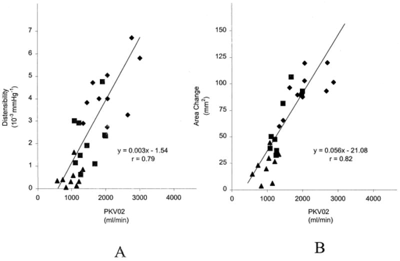
Metabolic exercise performance is directly related to aortic distensibility [A] and cross sectional area change [B] in patients with HFpEF, older-aged controls, and young healthy controls. From Hundley WG, Kitzman DW, Morgan TM, et al. Cardiac cycle-dependent changes in aortic area and distensibility are reduced in older patients with isolated diastolic heart failure and correlate with exercise intolerance. J Am Coll Cardiol. Sep 2001;38(3):796–802
In a noninvasive hemodynamic study, we compared subjects with HFpEF to matched controls with hypertensive LVH, and found that HFpEF patients displayed a markedly impaired ability to augment cardiac output with exercise, despite similar increases in preload36. The inability to augment cardiac output with stress correlated with chronotropic incompetence and an inability to vasodilate and drop systemic vascular resistance during exercise. Baseline blood pressure and vascular stiffness were similarly elevated in both groups, and only by measuring changes with exercise was the deficit in vascular reserve function observed. More recently, we found that among subjects with hypertensive heart disease referred to the cath lab for evaluation of dyspnea, acute exercise-induced increases in LV diastolic filling pressures were highest in subjects with the greatest exercise-induced increase in Ea and Ees, highlighting the importance of resting and exercise-induced stiffening in affecting exercise reserve54.
Therapeutic Strategies Targeting Stiffness
Ventricular-vascular stiffening can be treated with agents that acutely modulate ventricular systolic and diastolic performance, vascular smooth muscle tone, and endothelial function. Verapamil, which acutely reduces ventricular and vascular stiffness, improves exercise capacity in patients with HFpEF, hypertrophic cardiomyopathy, and elderly subjects with hypertension and hypertrophy55–57. A recent trial in older hypertensive patients found that verapamil led to significant reductions in ventricular and vascular stiffness and these changes were in turn associated with a 50% improvement in aerobic exercise performance57. Short term treatment with the angiotensin receptor antagonist losartan improves exercise performance in subjects with a hypertensive response to exercise as described above51, 52, while acute vasodilation with sodium nitroprusside did not share such beneficial effects58. Because isolated reduction in Ea results in greater decreases in blood pressure with less increase in stroke volume with a high baseline Ees (Fig 3), vasodilators that do not also treat ventricular stiffness will tend to have less benefit. Propranolol, which does decrease Ees, but also increases Ea, does not improve exercise performance59. Given the extent of chronotropic incompetence in HFpEF patients36, 60, drugs such as calcium channel antagonists and beta blockers require more study to compare offsetting effects on ventricular and arterial stiffness and heart rate, in addition to examining differences in acute versus chronic effects.
Many of the changes in ventricular-arterial stiffness in HFpEF are chronic, due to alterations in the material properties of the cardiovascular system25, 61. In the largest randomized trial of HF with near-normal EF, there was a borderline-significant treatment effect driven by a reduction in hospitalizations for HF62. However, this trial enrolled subjects with HF and EF>40%, and many would consider those with EF<50–55% to have some element of systolic dysfunction26,27. ACE-inhibitors, which also reduce vascular stiffness, may be useful in HFpEF63. Other treatments under evaluation include aldosterone64, 65, TGF-β66, and chymase antagonists67. A large-scale randomized, NIH-funded trial is currently underway testing the efficacy of the aldosterone antagonist spironolactone in HFpEF.
Ventricular hypertrophy is commonly seen in HFpEF, and the increase in stiffening and concentric remodeling seen with hypertensive heart disease commonly result in increased Ees, thus hypertrophy may be a novel therapeutic target in this disorder and those at risk. Controlling blood pressure and afterload is the most obvious way to ameliorate hypertrophy, but strategies specifically targeting concentric remodeling may be synergistically useful as well. Among the panoply of candidate small molecules involved in hypertrophic signaling is rho kinase, a key downstream effecter of Gq-protein coupled angiotensin-II signaling24, 68. Rho kinase increases vascular smooth muscle cell tone, and the rho kinase inhibitor fasudil is being evaluated as an anti-anginal drug. Fasudil also attenuates Ang-II-induced cardiac hypertrophy, thus therapies interfering with rho kinase may prove synergistically useful related its vasodilatory and anti-hypertrophic properties69, 70. HMG CoA-reductase inhibitors (statins) have recently been shown to be associated with improved survival in a retrospective analysis of patients with HFpEF, and intriguingly, are known to also inhibit Rho kinase activation70, 71. We recently demonstrated that the phosphodiesterase 5 inhibitor sildenafil prevents the hypertrophic response to pressure overload in mice72. In humans, sildenafil suppresses acute β-adrenergic stimulated contractility73. The NIH-sponsored RELAX trial will soon begin enrollment, studying whether chronic PDE5 inhibition with sildenafil can improve exercise performance and reduce ventricular hypertrophy and vascular stiffening in HFpEF patients.
As clinically-based pathophysiologic research continues to identify mechanisms whereby ventricular-arterial stiffening impairs cardiovascular function, and basic research defines the key cellular players involved in promoting ventriculoarterial stiffness, remodeling, and hypertrophy, we will increasingly be able to better treat patients with HFpEF by targeting these highly complex coexisting, and highly-integrated pathophysiologic pathways related to ventriculoarterial stiffening.
Footnotes
Publisher's Disclaimer: This is a PDF file of an unedited manuscript that has been accepted for publication. As a service to our customers we are providing this early version of the manuscript. The manuscript will undergo copyediting, typesetting, and review of the resulting proof before it is published in its final citable form. Please note that during the production process errors may be discovered which could affect the content, and all legal disclaimers that apply to the journal pertain.
References
- 1.Suga H, Sagawa K. Instantaneous pressure-volume relationships and their ratio in the excised, supported canine left ventricle. Circ Res Jul. 1974;35(1):117–126. doi: 10.1161/01.res.35.1.117. [DOI] [PubMed] [Google Scholar]
- 2.Kass DA, Kelly RP. Ventriculo-arterial coupling: concepts, assumptions, and applications. Ann Biomed Eng. 1992;20(1):41–62. doi: 10.1007/BF02368505. [DOI] [PubMed] [Google Scholar]
- 3.Milnor WR. Arterial impedance as ventricular afterload. Circ Res May. 1975;36(5):565–570. doi: 10.1161/01.res.36.5.565. [DOI] [PubMed] [Google Scholar]
- 4.Murgo JP, Westerhof N, Giolma JP, Altobelli SA. Aortic input impedance in normal man: relationship to pressure wave forms. Circulation Jul. 1980;62(1):105–116. doi: 10.1161/01.cir.62.1.105. [DOI] [PubMed] [Google Scholar]
- 5.Sunagawa K, Maughan WL, Burkhoff D, Sagawa K. Left ventricular interaction with arterial load studied in isolated canine ventricle. Am J Physiol Nov. 1983;245(5 Pt 1):H773–780. doi: 10.1152/ajpheart.1983.245.5.H773. [DOI] [PubMed] [Google Scholar]
- 6.Sunagawa K, Maughan WL, Sagawa K. Optimal arterial resistance for the maximal stroke work studied in isolated canine left ventricle. Circ Res Apr. 1985;56(4):586–595. doi: 10.1161/01.res.56.4.586. [DOI] [PubMed] [Google Scholar]
- 7.Kelly RP, Ting CT, Yang TM, et al. Effective arterial elastance as index of arterial vascular load in humans. Circulation Aug. 1992;86(2):513–521. doi: 10.1161/01.cir.86.2.513. [DOI] [PubMed] [Google Scholar]
- 8.Asanoi H, Sasayama S, Kameyama T. Ventriculoarterial coupling in normal and failing heart in humans. Circ Res Aug. 1989;65(2):483–493. doi: 10.1161/01.res.65.2.483. [DOI] [PubMed] [Google Scholar]
- 9.De Tombe PP, Jones S, Burkhoff D, Hunter WC, Kass DA. Ventricular stroke work and efficiency both remain nearly optimal despite altered vascular loading. Am J Physiol Jun. 1993;264(6 Pt 2):H1817–1824. doi: 10.1152/ajpheart.1993.264.6.H1817. [DOI] [PubMed] [Google Scholar]
- 10.Redfield MM, Jacobsen SJ, Borlaug BA, Rodeheffer RJ, Kass DA. Age- and gender-related ventricular-vascular stiffening: a community-based study. Circulation. 2005 Oct 11;112(15):2254–2262. doi: 10.1161/CIRCULATIONAHA.105.541078. [DOI] [PubMed] [Google Scholar]
- 11.Najjar SS, Schulman SP, Gerstenblith G, et al. Age and gender affect ventricular-vascular coupling during aerobic exercise. J Am Coll Cardiol. 2004 Aug 4;44(3):611–617. doi: 10.1016/j.jacc.2004.04.041. [DOI] [PubMed] [Google Scholar]
- 12.Avolio AP, Chen SG, Wang RP, Zhang CL, Li MF, O'Rourke MF. Effects of aging on changing arterial compliance and left ventricular load in a northern Chinese urban community. Circulation Jul. 1983;68(1):50–58. doi: 10.1161/01.cir.68.1.50. [DOI] [PubMed] [Google Scholar]
- 13.Chen CH, Nakayama M, Nevo E, Fetics BJ, Maughan WL, Kass DA. Coupled systolic-ventricular and vascular stiffening with age: implications for pressure regulation and cardiac reserve in the elderly. J Am Coll Cardiol Nov. 1998;32(5):1221–1227. doi: 10.1016/s0735-1097(98)00374-x. [DOI] [PubMed] [Google Scholar]
- 14.Nichols WW, O'Rourke MF, Avolio AP, et al. Effects of age on ventricular-vascular coupling. Am J Cardiol. 1985 Apr 15;55(9):1179–1184. doi: 10.1016/0002-9149(85)90659-9. [DOI] [PubMed] [Google Scholar]
- 15.Kawaguchi M, Hay I, Fetics B, Kass DA. Combined ventricular systolic and arterial stiffening in patients with heart failure and preserved ejection fraction: implications for systolic and diastolic reserve limitations. Circulation. 2003 Feb 11;107(5):714–720. doi: 10.1161/01.cir.0000048123.22359.a0. [DOI] [PubMed] [Google Scholar]
- 16.Nichols WW, O'Rourke MF, Avolio AP, Yaginuma T, Pepine CJ, Conti CR. Ventricular/vascular interaction in patients with mild systemic hypertension and normal peripheral resistance. Circulation Sep. 1986;74(3):455–462. doi: 10.1161/01.cir.74.3.455. [DOI] [PubMed] [Google Scholar]
- 17.Segers P, Stergiopulos N, Westerhof N. Relation of effective arterial elastance to arterial system properties. Am J Physiol Heart Circ Physiol Mar. 2002;282(3):H1041–1046. doi: 10.1152/ajpheart.00764.2001. [DOI] [PubMed] [Google Scholar]
- 18.Chemla D, Antony I, Lecarpentier Y, Nitenberg A. Contribution of systemic vascular resistance and total arterial compliance to effective arterial elastance in humans. Am J Physiol Heart Circ Physiol Aug. 2003;285(2):H614–620. doi: 10.1152/ajpheart.00823.2002. [DOI] [PubMed] [Google Scholar]
- 19.Kelly R, Hayward C, Avolio A, O'Rourke M. Noninvasive determination of age-related changes in the human arterial pulse. Circulation Dec. 1989;80(6):1652–1659. doi: 10.1161/01.cir.80.6.1652. [DOI] [PubMed] [Google Scholar]
- 20.Borlaug BA, Melenovsky V, Redfield MM, et al. The Impact of Arterial Load and Loading Sequence On Left Ventricular Tissue Velocities in Humans. J Am Coll Card. 2007 Oct 16; doi: 10.1016/j.jacc.2007.07.032. [DOI] [PubMed] [Google Scholar]
- 21.Melenovsky V, Borlaug BA, Rosen B, et al. Cardiovascular features of heart failure with preserved ejection fraction versus nonfailing hypertensive left ventricular hypertrophy in the urban Baltimore community: the role of atrial remodeling/dysfunction. J Am Coll Cardiol. 2007 Jan 16;49(2):198–207. doi: 10.1016/j.jacc.2006.08.050. [DOI] [PubMed] [Google Scholar]
- 22.Lam CS, Roger VL, Rodeheffer RJ, et al. Cardiac structure and ventricular-vascular function in persons with heart failure and preserved ejection fraction from Olmsted County, Minnesota. Circulation. 2007 Apr 17;115(15):1982–1990. doi: 10.1161/CIRCULATIONAHA.106.659763. [DOI] [PMC free article] [PubMed] [Google Scholar]
- 23.Zile MR, Baicu CF, Gaasch WH. Diastolic heart failure--abnormalities in active relaxation and passive stiffness of the left ventricle. N Engl J Med. 2004 May 6;350(19):1953–1959. doi: 10.1056/NEJMoa032566. [DOI] [PubMed] [Google Scholar]
- 24.Borlaug BA, Kass DA. Mechanisms of diastolic dysfunction in heart failure. Trends Cardiovasc Med. 2006 Nov;16(8):273–279. doi: 10.1016/j.tcm.2006.05.003. [DOI] [PubMed] [Google Scholar]
- 25.Zieman SJ, Melenovsky V, Kass DA. Mechanisms, pathophysiology, and therapy of arterial stiffness. Arterioscler Thromb Vasc Biol. 2005 May;25(5):932–943. doi: 10.1161/01.ATV.0000160548.78317.29. [DOI] [PubMed] [Google Scholar]
- 26.Owan TE, Redfield MM. Epidemiology of diastolic heart failure. Prog Cardiovasc Dis. 2005 Mar–Apr;47(5):320–332. doi: 10.1016/j.pcad.2005.02.010. [DOI] [PubMed] [Google Scholar]
- 27.Klapholz M, Maurer M, Lowe AM, et al. Hospitalization for heart failure in the presence of a normal left ventricular ejection fraction: results of the New York Heart Failure Registry. J Am Coll Cardiol. 2004 Apr 21;43(8):1432–1438. doi: 10.1016/j.jacc.2003.11.040. [DOI] [PubMed] [Google Scholar]
- 28.Borbely A, van der Velden J, Papp Z, et al. Cardiomyocyte stiffness in diastolic heart failure. Circulation. 2005 Feb 15;111(6):774–781. doi: 10.1161/01.CIR.0000155257.33485.6D. [DOI] [PubMed] [Google Scholar]
- 29.van Heerebeek L, Borbely A, Niessen HW, et al. Myocardial structure and function differ in systolic and diastolic heart failure. Circulation. 2006 Apr 25;113(16):1966–1973. doi: 10.1161/CIRCULATIONAHA.105.587519. [DOI] [PubMed] [Google Scholar]
- 30.Dauterman K, Pak PH, Maughan WL, et al. Contribution of external forces to left ventricular diastolic pressure. Implications for the clinical use of the Starling law. Ann Intern Med. 1995 May 15;122(10):737–742. doi: 10.7326/0003-4819-122-10-199505150-00001. [DOI] [PubMed] [Google Scholar]
- 31.Gandhi SK, Powers JC, Nomeir AM, et al. The pathogenesis of acute pulmonary edema associated with hypertension. N Engl J Med. 2001 Jan 4;344(1):17–22. doi: 10.1056/NEJM200101043440103. [DOI] [PubMed] [Google Scholar]
- 32.Carroll JD, Carroll EP, Feldman T, et al. Sex-associated differences in left ventricular function in aortic stenosis of the elderly. Circulation. 1992 Oct;86(4):1099–1107. doi: 10.1161/01.cir.86.4.1099. [DOI] [PubMed] [Google Scholar]
- 33.Villari B, Campbell SE, Schneider J, Vassalli G, Chiariello M, Hess OM. Sex-dependent differences in left ventricular function and structure in chronic pressure overload. Eur Heart J. 1995 Oct;16(10):1410–1419. doi: 10.1093/oxfordjournals.eurheartj.a060749. [DOI] [PubMed] [Google Scholar]
- 34.Kass DA, Maughan WL. From 'Emax' to pressure-volume relations: a broader view. Circulation. 1988 Jun;77(6):1203–1212. doi: 10.1161/01.cir.77.6.1203. [DOI] [PubMed] [Google Scholar]
- 35.Franciosa JA, Leddy CL, Wilen M, Schwartz DE. Relation between hemodynamic and ventilatory responses in determining exercise capacity in severe congestive heart failure. Am J Cardiol. 1984 Jan 1;53(1):127–134. doi: 10.1016/0002-9149(84)90696-9. [DOI] [PubMed] [Google Scholar]
- 36.Borlaug BA, Melenovsky V, Russell SD, et al. Impaired chronotropic and vasodilator reserves limit exercise capacity in patients with heart failure and a preserved ejection fraction. Circulation. 2006 Nov 14;114(20):2138–2147. doi: 10.1161/CIRCULATIONAHA.106.632745. [DOI] [PubMed] [Google Scholar]
- 37.Gillebert TC, Leite-Moreira AF, De Hert SG. Load dependent diastolic dysfunction in heart failure. Heart Fail Rev. 2000 Dec;5(4):345–355. doi: 10.1023/a:1026563313952. [DOI] [PubMed] [Google Scholar]
- 38.Gillebert TC, Lew WY. Influence of systolic pressure profile on rate of left ventricular pressure fall. Am J Physiol. 1991 Sep;261(3 Pt 2):H805–813. doi: 10.1152/ajpheart.1991.261.3.H805. [DOI] [PubMed] [Google Scholar]
- 39.Leite-Moreira AF, Correia-Pinto J, Gillebert TC. Afterload induced changes in myocardial relaxation: a mechanism for diastolic dysfunction. Cardiovasc Res. 1999 Aug 1;43(2):344–353. doi: 10.1016/s0008-6363(99)00099-1. [DOI] [PubMed] [Google Scholar]
- 40.Takimoto E, Soergel DG, Janssen PM, Stull LB, Kass DA, Murphy AM. Frequency- and afterload-dependent cardiac modulation in vivo by troponin I with constitutively active protein kinase A phosphorylation sites. Circ Res. 2004 Mar 5;94(4):496–504. doi: 10.1161/01.RES.0000117307.57798.F5. [DOI] [PubMed] [Google Scholar]
- 41.Bilchick KC, Duncan JG, Ravi R, et al. Heart failure-associated alterations in troponin I phosphorylation impair ventricular relaxation-afterload and force-frequency responses and systolic function. Am J Physiol Heart Circ Physiol. 2006 Aug 25; doi: 10.1152/ajpheart.00283.2006. [DOI] [PubMed] [Google Scholar]
- 42.Brutsaert DL, Sys SU. Relaxation and diastole of the heart. Physiol Rev. 1989 Oct;69(4):1228–1315. doi: 10.1152/physrev.1989.69.4.1228. [DOI] [PubMed] [Google Scholar]
- 43.Solomon SD, Janardhanan R, Verma A, et al. Effect of angiotensin receptor blockade and antihypertensive drugs on diastolic function in patients with hypertension and diastolic dysfunction: a randomised trial. Lancet. 2007 Jun 23;369(9579):2079–2087. doi: 10.1016/S0140-6736(07)60980-5. [DOI] [PubMed] [Google Scholar]
- 44.Gilbert JC, Glantz SA. Determinants of left ventricular filling and of the diastolic pressure-volume relation. Circ Res. 1989 May;64(5):827–852. doi: 10.1161/01.res.64.5.827. [DOI] [PubMed] [Google Scholar]
- 45.Kass DA, Wolff MR, Ting CT, et al. Diastolic compliance of hypertrophied ventricle is not acutely altered by pharmacologic agents influencing active processes. Ann Intern Med. 1993 Sep 15;119(6):466–473. doi: 10.7326/0003-4819-119-6-199309150-00004. [DOI] [PubMed] [Google Scholar]
- 46.Leite-Moreira AF, Correia-Pinto J. Load as an acute determinant of end-diastolic pressure-volume relation. Am J Physiol Heart Circ Physiol. 2001 Jan;280(1):H51–59. doi: 10.1152/ajpheart.2001.280.1.H51. [DOI] [PubMed] [Google Scholar]
- 47.Shapiro BP, Lam CS, Patel JB, et al. Acute and Chronic Ventricular-Arterial Coupling in Systole and Diastole. Insights From an Elderly Hypertensive Model. Hypertension. 2007 Jul 9; doi: 10.1161/HYPERTENSIONAHA.107.090092. [DOI] [PubMed] [Google Scholar]
- 48.Saeki A, Recchia F, Kass DA. systolic flow augmentation in hearts ejecting into a model of stiff aging vasculature. Influence on myocardial perfusion-demand balance. Circ Res. 1995 Jan;76(1):132–141. doi: 10.1161/01.res.76.1.132. [DOI] [PubMed] [Google Scholar]
- 49.Kass DA, Saeki A, Tunin RS, Recchia FA. Adverse influence of systemic vascular stiffening on cardiac dysfunction and adaptation to acute coronary occlusion. Circulation. 1996 Apr 15;93(8):1533–1541. doi: 10.1161/01.cir.93.8.1533. [DOI] [PubMed] [Google Scholar]
- 50.Li M, Chiou KR, Bugayenko A, Irani K, Kass DA. Reduced wall compliance suppresses Akt-dependent apoptosis protection stimulated by pulse perfusion. Circ Res. 2005 Sep 16;97(6):587–595. doi: 10.1161/01.RES.0000181432.73920.b1. [DOI] [PubMed] [Google Scholar]
- 51.Warner JG, Jr, Metzger DC, Kitzman DW, Wesley DJ, Little WC. Losartan improves exercise tolerance in patients with diastolic dysfunction and a hypertensive response to exercise. J Am Coll Cardiol. 1999 May;33(6):1567–1572. doi: 10.1016/s0735-1097(99)00048-0. [DOI] [PubMed] [Google Scholar]
- 52.Little WC, Zile MR, Klein A, Appleton CP, Kitzman DW, Wesley-Farrington DJ. Effect of losartan and hydrochlorothiazide on exercise tolerance in exertional hypertension and left ventricular diastolic dysfunction. Am J Cardiol. 2006 Aug 1;98(3):383–385. doi: 10.1016/j.amjcard.2006.01.106. [DOI] [PubMed] [Google Scholar]
- 53.Hundley WG, Kitzman DW, Morgan TM, et al. Cardiac cycle-dependent changes in aortic area and distensibility are reduced in older patients with isolated diastolic heart failure and correlate with exercise intolerance. J Am Coll Cardiol. 2001 Sep;38(3):796–802. doi: 10.1016/s0735-1097(01)01447-4. [DOI] [PubMed] [Google Scholar]
- 54.Jaber WA, Nishimura RA, Borlaug BA, et al. Determination of the Mechanism of Elevated Left Ventricular Filling Pressures with Exercise: A Simultaneous Echocardiographic-Catheterization Study. Circulation. 2007:1756. (abs) [Google Scholar]
- 55.Bonow RO, Dilsizian V, Rosing DR, Maron BJ, Bacharach SL, Green MV. Verapamil-induced improvement in left ventricular diastolic filling and increased exercise tolerance in patients with hypertrophic cardiomyopathy: short- and long-term effects. Circulation. 1985 Oct;72(4):853–864. doi: 10.1161/01.cir.72.4.853. [DOI] [PubMed] [Google Scholar]
- 56.Setaro JF, Zaret BL, Schulman DS, Black HR, Soufer R. Usefulness of verapamil for congestive heart failure associated with abnormal left ventricular diastolic filling and normal left ventricular systolic performance. Am J Cardiol. 1990 Oct 15;66(12):981–986. doi: 10.1016/0002-9149(90)90937-v. [DOI] [PubMed] [Google Scholar]
- 57.Chen CH, Nakayama M, Talbot M, et al. Verapamil acutely reduces ventricular-vascular stiffening and improves aerobic exercise performance in elderly individuals. J Am Coll Cardiol. 1999 May;33(6):1602–1609. doi: 10.1016/s0735-1097(99)00052-2. [DOI] [PubMed] [Google Scholar]
- 58.Nussbacher A, Gerstenblith G, O'Connor FC, et al. Hemodynamic effects of unloading the old heart. Am J Physiol. 1999 Nov;277(5 Pt 2):H1863–1871. doi: 10.1152/ajpheart.1999.277.5.H1863. [DOI] [PubMed] [Google Scholar]
- 59.Fleg JL, Schulman S, O'Connor F, et al. Effects of acute beta-adrenergic receptor blockade on age-associated changes in cardiovascular performance during dynamic exercise. Circulation. 1994 Nov;90(5):2333–2341. doi: 10.1161/01.cir.90.5.2333. [DOI] [PubMed] [Google Scholar]
- 60.Brubaker PH, Joo KC, Stewart KP, Fray B, Moore B, Kitzman DW. Chronotropic incompetence and its contribution to exercise intolerance in older heart failure patients. J Cardiopulm Rehabil. 2006 Mar–Apr;26(2):86–89. doi: 10.1097/00008483-200603000-00007. [DOI] [PubMed] [Google Scholar]
- 61.Kass DA. Ventricular arterial stiffening: integrating the pathophysiology. Hypertension. 2005 Jul;46(1):185–193. doi: 10.1161/01.HYP.0000168053.34306.d4. [DOI] [PubMed] [Google Scholar]
- 62.Yusuf S, Pfeffer MA, Swedberg K, et al. Effects of candesartan in patients with chronic heart failure and preserved left-ventricular ejection fraction: the CHARM-Preserved Trial. Lancet. 2003 Sep 6;362(9386):777–781. doi: 10.1016/S0140-6736(03)14285-7. [DOI] [PubMed] [Google Scholar]
- 63.Cleland JG, Tendera M, Adamus J, Freemantle N, Polonski L, Taylor J. The perindopril in elderly people with chronic heart failure (PEP-CHF) study. Eur Heart J. 2006 Sep 8; doi: 10.1093/eurheartj/ehl250. [DOI] [PubMed] [Google Scholar]
- 64.Pitt B, Reichek N, Willenbrock R, et al. Effects of eplerenone, enalapril, and eplerenone/enalapril in patients with essential hypertension and left ventricular hypertrophy: the 4E-left ventricular hypertrophy study. Circulation. 2003 Oct 14;108(15):1831–1838. doi: 10.1161/01.CIR.0000091405.00772.6E. [DOI] [PubMed] [Google Scholar]
- 65.Pitt B, Zannad F, Remme WJ, et al. The effect of spironolactone on morbidity and mortality in patients with severe heart failure. Randomized Aldactone Evaluation Study Investigators. N Engl J Med. 1999 Sep 2;341(10):709–717. doi: 10.1056/NEJM199909023411001. [DOI] [PubMed] [Google Scholar]
- 66.Kuwahara F, Kai H, Tokuda K, et al. Transforming growth factor-beta function blocking prevents myocardial fibrosis and diastolic dysfunction in pressure-overloaded rats. Circulation. 2002 Jul 2;106(1):130–135. doi: 10.1161/01.cir.0000020689.12472.e0. [DOI] [PubMed] [Google Scholar]
- 67.Matsumoto T, Wada A, Tsutamoto T, Ohnishi M, Isono T, Kinoshita M. Chymase inhibition prevents cardiac fibrosis and improves diastolic dysfunction in the progression of heart failure. Circulation. 2003 May 27;107(20):2555–2558. doi: 10.1161/01.CIR.0000074041.81728.79. [DOI] [PubMed] [Google Scholar]
- 68.Lai A, Frishman WH. Rho-kinase inhibition in the therapy of cardiovascular disease. Cardiol Rev. 2005 Nov–Dec;13(6):285–292. doi: 10.1097/01.crd.0000138079.91392.37. [DOI] [PubMed] [Google Scholar]
- 69.Higashi M, Shimokawa H, Hattori T, et al. Long-term inhibition of Rho-kinase suppresses angiotensin II-induced cardiovascular hypertrophy in rats in vivo: effect on endothelial NAD(P)H oxidase system. Circ Res. 2003 Oct 17;93(8):767–775. doi: 10.1161/01.RES.0000096650.91688.28. [DOI] [PubMed] [Google Scholar]
- 70.Morikawa-Futamatsu K, Adachi S, Maejima Y, et al. HMG-CoA reductase inhibitor fluvastatin prevents angiotensin II-induced cardiac hypertrophy via Rho kinase and inhibition of cyclin D1. Life Sci. 2006 Aug 29;79(14):1380–1390. doi: 10.1016/j.lfs.2006.04.005. [DOI] [PubMed] [Google Scholar]
- 71.Fukuta H, Sane DC, Brucks S, Little WC. Statin therapy may be associated with lower mortality in patients with diastolic heart failure: a preliminary report. Circulation. 2005 Jul 19;112(3):357–363. doi: 10.1161/CIRCULATIONAHA.104.519876. [DOI] [PubMed] [Google Scholar]
- 72.Takimoto E, Champion HC, Li M, et al. Chronic inhibition of cyclic GMP phosphodiesterase 5A prevents and reverses cardiac hypertrophy. Nat Med. 2005 Feb;11(2):214–222. doi: 10.1038/nm1175. [DOI] [PubMed] [Google Scholar]
- 73.Borlaug BA, Melenovsky V, Marhin T, Fitzgerald P, Kass DA. Sildenafil inhibits beta-adrenergic-stimulated cardiac contractility in humans. Circulation. 2005 Oct 25;112(17):2642–2649. doi: 10.1161/CIRCULATIONAHA.105.540500. [DOI] [PubMed] [Google Scholar]
- 74.Kraemer MD, Kubo SH, Rector TS, Brunsvold N, Bank AJ. Pulmonary and peripheral vascular factors are important determinants of peak exercise oxygen uptake in patients with heart failure. J Am Coll Cardiol. 1993 Mar 1;21(3):641–648. doi: 10.1016/0735-1097(93)90096-j. [DOI] [PubMed] [Google Scholar]