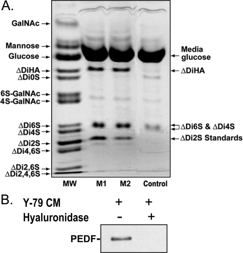
FIGURE 1.
PEDF association to hyaluronan secreted by human retinoblastoma Y-79 cells. A, human retinoblastoma Y-79 cells (1.25 × 10⁵ cells/ml) were cultured in serum-free medium for 16 h, and then the culture conditioned media were collected and concentrated 10-fold before being subjected to fluorophore-assisted carbohydrate electrophoresis. The fluorogram shows the separation of saccharides recovered from Y-79-conditioned media from two different batches of Y-79 cell cultures (M1 and M2). The image shown depicts oversaturated pixel intensity for the major derivatized components to allow visualization of less abundant components. The lane marked MW shows the resolution of a standard mixture of 13 AMAC-derivatized saccharides. The Control lane shows a derivatized medium sample not conditioned by human retinoblastoma Y-79 cells for background corrections. ΔDi2S standards at 62.5 and 31.5 pmol (as determined by hexuronic acid analysis) were added to lanes marked M1 and M2, respectively, for the quantification of resolved disaccharides. B, PEDF coprecipitates with glycosaminoglycans secreted into the Y-79-conditioned media. Serum-free medium from human retinoblastoma cell cultures (1.25 × 10⁶ cells/ml) maintained at 37 °C for 16 h was concentrated (25-fold), and 500-μl aliquots were either left untreated or treated with Streptomyces hyluronidase (10 turbidity reducing units/ml) for 10 min at 37 °C. Recombinant human PEDF (20 μg/ml) and BSA (100 μg/ml) were added, and the mixtures were incubated at 4 °C for 120 min. Glycosaminoglycans were precipitated with CPC. A Western blot of the precipitates was immunostained with Ab-rPEDF, and is shown. Treatments are indicated at the top of each lane.

