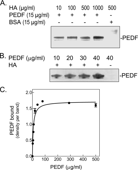
FIGURE 2.
Direct binding of PEDF to HA. Purified recombinant human PEDF and highly purified hyaluronan (Healon®) were incubated at 4 °C for 1 h. HA was precipitated with CPC, and the pellets were resuspended in SDS-PAGE sample buffer and applied to 10–20% polyacrylamide gels for detection of PEDF. A, binding reactions were performed with recombinant human PEDF and increasing concentrations of HA (indicated at the top of each lane) in 200 μl of buffer H (20 mm sodium phosphate, pH 6.5, 20 mm NaCl, and 10% glycerol). BSA was the negative control. Proteins in the precipitates were resolved by SDS-PAGE and visualized with Silver Stain. A photograph of a stained gel is shown. B, binding reactions were performed with 100 μg/ml HA and increasing concentrations of PEDF (indicated at the top of each lane) in 500 μl of Dulbecco's modified Eagle's medium plus 100 μg/ml BSA. PEDF in the precipitates was detected by Western blotting with antiserum Ab-rPEDF. C, concentration curve of PEDF binding to HA. Binding reactions were performed with 200 μg/ml HA and increasing PEDF concentrations in 200 μl of 150 mm NaCl in Buffer S (20 mm sodium phosphate, pH 6.4, 10% glycerol, 1 mm dithiothreitol) plus 100 μg/ml BSA (carrier protein). One-third of each reaction mixture was resolved by SDS-PAGE followed by silver staining, and two remaining thirds were analyzed by Western blotting with Ab-rPEDF. The density of each PEDF band was determined from scanned images using Stratagene Eagle Eye II. Data were analyzed by non-linear regression and one-site binding using a GraphPad Prism version 3.0 software program (plot shown). The best-fit values obtained from data combined from silver-stained gels and Western transfers were Bmax = 1.723 bound PEDF, and KD = 7.027 μg/ml PEDF (= 140.54 nm), with an R2 = 0.9715.

