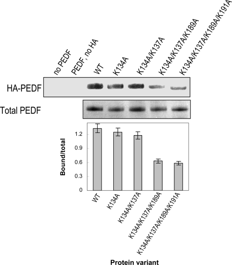
FIGURE 6.
Binding to HA of PEDF modified on putative exposed lysines. Altered PEDF proteins were purified and concentrated. Purified altered PEDF proteins and HA were mixed in buffer H and incubated at 4 °C for 1 h. Bound protein was coprecipitated with CPC. Western blot of CPC precipitates immunostained with anti-PEDF is shown at the top (HA-PEDF). SDS-PAGE of purified PEDF altered proteins stained with Coomassie Brilliant Blue is shown at the bottom (Total PEDF). A plot of bound PEDF quantification was performed as in Fig. 5C.

